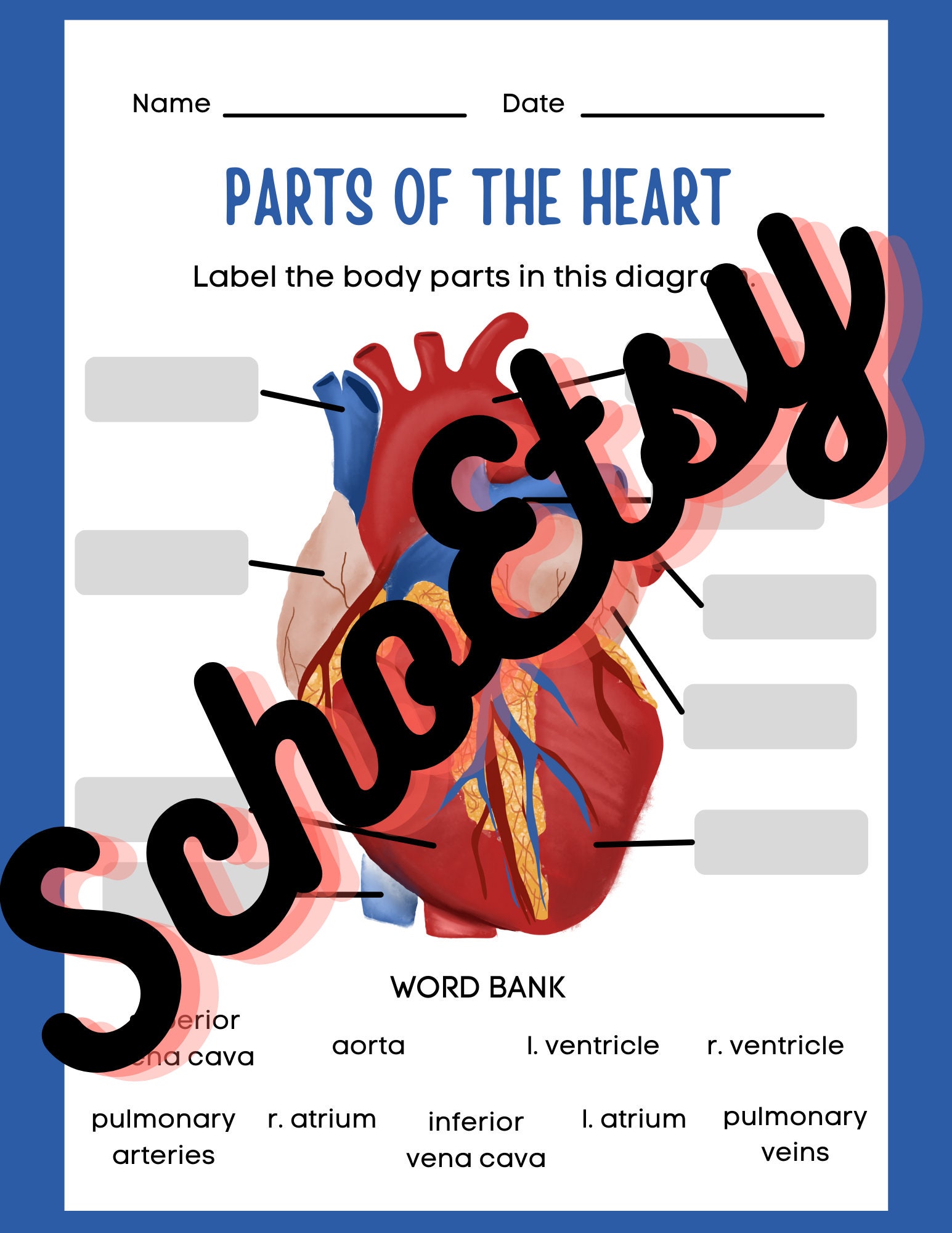
43 label heart parts
Labelling the heart — Science Learning Hub identify the main parts of a heart describe the functions of the different parts of the heart. This online interactive supports students to identify the different parts of the heart and the role they have in blood circulation. It can be used as both a formative and summative tool for learning. Topics Concepts Citizen science Teacher PLD Glossary Diagrams, quizzes and worksheets of the heart | Kenhub Labeled heart diagrams Take a look at our labeled heart diagrams (see below) to get an overview of all of the parts of the heart. Once you're feeling confident, you can test yourself using the unlabeled diagrams of the parts of the heart below. Labeled heart diagram showing the heart from anterior Unlabeled heart diagrams (free download!)
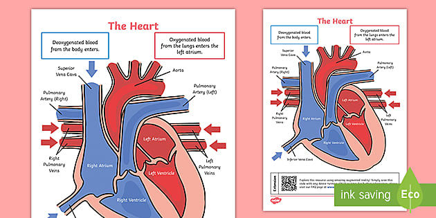
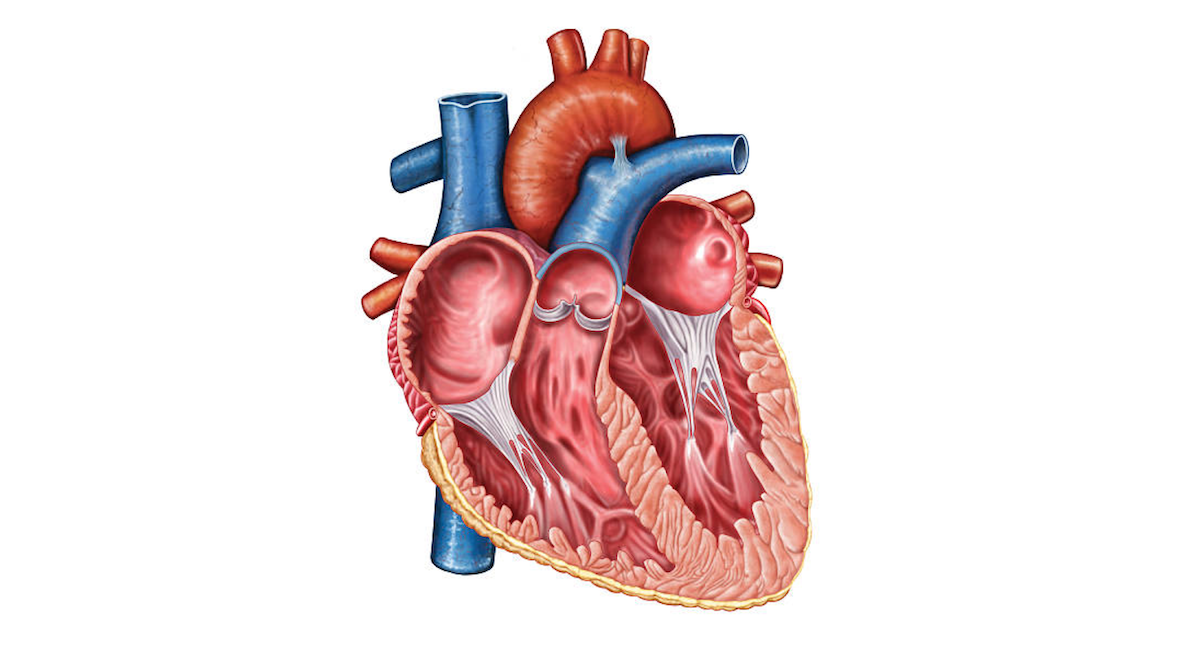
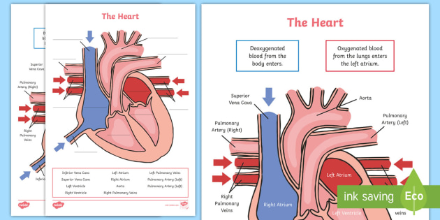
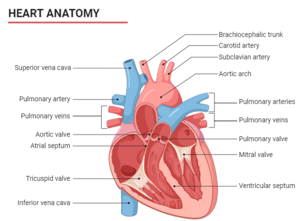
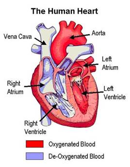
A Labeled Diagram of the Human Heart You Really Need to See The human heart, comprises four chambers: right atrium, left atrium, right ventricle and left ventricle. The two upper chambers are called the left and the right atria, and the two lower chambers are known as the left and the right ventricles. The two atria and ventricles are separated from each other by a muscle wall called 'septum'.
Label heart parts
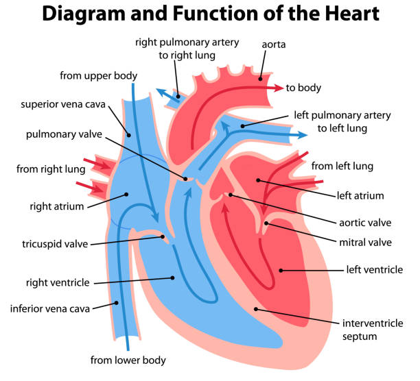
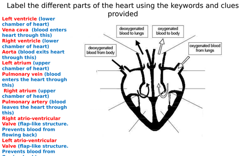
Heart: Anatomy and Function - Cleveland Clinic The parts of your heart are like the parts of a house. Your heart has: Walls. Chambers (rooms). Valves (doors). Blood vessels (plumbing). Electrical conduction system (electricity). Heart walls Your heart walls are the muscles that contract (squeeze) and relax to send blood throughout your body. Label the heart — Science Learning Hub Label the heart Interactive Add to collection In this interactive, you can label parts of the human heart. Drag and drop the text labels onto the boxes next to the diagram. Selecting or hovering over a box will highlight each area in the diagram. semilunar valve aorta left ventricle right ventricle left atrium vena cava pulmonary artery Human Heart - Diagram and Anatomy of the Heart - Innerbody The heart contains 4 chambers: the right atrium, left atrium, right ventricle, and left ventricle. The atria are smaller than the ventricles and have thinner, less muscular walls than the ventricles. The atria act as receiving chambers for blood, so they are connected to the veins that carry blood to the heart.
Label heart parts. Label Parts Of A Heart - Label The Heart Diagram Quizlet The heart is a muscular organ about the size of a fist, located just behind and slightly left of the breastbone. Aortic valve left atrium left ventricle mitral valve pulmonary valve right atrium. The more healthy your heart is, the longer the . Structure of the heart use the word bank to label the parts of the heart. Picture of the Heart - WebMD The heart is a muscular organ about the size of a fist, located just behind and slightly left of the breastbone. The heart pumps blood through the network of arteries and veins called the... Label the heart - Teaching resources - Wordwall KS4 Y10 Y11 Biology Science. 10X Label the Heart diagram Labelled diagram. by Kpatel1. Label The Diagram of The Heart Labelled diagram. by Paulrobertedwards. Label The Diagram of The Heart Labelled diagram. by Eadams4. KS2 Science Living things. Y5 Label The Diagram of The Heart Labelled diagram. Heart Labeling Quiz: How Much You Know About Heart Labeling? Here is a Heart labeling quiz for you. The human heart is a vital organ for every human. The more healthy your heart is, the longer the chances you have of surviving, so you better take care of it. Take the following quiz to know how much you know about your heart. Questions and Answers 1. What is #1? 2. What is #2? 3. What is #3? 4. What is #4?
Label the Heart Diagram | Quizlet The Respiratory System (Label) 9 terms Diagram. steve_murks Teacher. Label the Heart. 13 terms Images. BootstrapTeacher Teacher. Parts Of The Human Heart - Science Trends The parts of the human heart can be broken down into four chambers, muscular walls, vessels, and a conductive system. The two upper chambers are called the atria, with lower parts called ventricles. These all work together to make up the vital function of your heart. Everybody knows that the human heart is the essential organ in our bodies. Heart | Structure, Function, Diagram, Anatomy, & Facts heart, organ that serves as a pump to circulate the blood. It may be a straight tube, as in spiders and annelid worms, or a somewhat more elaborate structure with one or more receiving chambers (atria) and a main pumping chamber (ventricle), as in mollusks. In fishes the heart is a folded tube, with three or four enlarged areas that correspond to the chambers in the mammalian heart. Diagram of Human Heart and Blood Circulation in It The wall of the heart has three different layers, such as the Myocardium, the Epicardium, and the Endocardium. Here's more about these three layers. Epicardium The outermost layer of your heart wall is called the epicardium, which is basically a very thin layer of serous membrane.
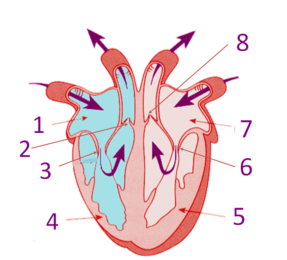
Heart Diagram with Labels and Detailed Explanation - BYJUS The heart is made up of four chambers: The upper two chambers of the heart are called auricles. The lower two chambers of the heart are called ventricles. The heart wall is made up of three layers: The outer layer of the heart wall is called epicardium. The middle layer of the heart wall is called myocardium. Human Anatomy: Label the Heart Worksheets in 3 Differentiated Levels Label the Heart Worksheets for Biology and Human Anatomy Study The Level 1 worksheet asks students to label the following parts of the heart: Aorta Vena Cava Right & Left Auricles Right & Left Ventricles The Level 2 worksheet asks students to label the above parts, plus the following: Superior and Inferior Vena Cava Aortic Arch Human Heart Diagram Labeled - Science Trends The heart has four different chambers: the left and right ventricles and the left and right atriums. The chambers of the heart and the valves that regulate blood flow to them are considered the plumbing of the heart. The left ventricle and left atrium make up the left heart while the right ventricle and right atrium make up the right heart. A Diagram of the Heart and Its Functioning Explained in Detail The heart blood flow diagram (flowchart) given below will help you to understand the pathway of blood through the heart.Initial five points denotes impure or deoxygenated blood and the last five points denotes pure or oxygenated blood. 1.Different Parts of the Body. ↓. 2.Major Veins.
Heart: Anatomy and Function
Label Heart Anatomy Diagram Printout - EnchantedLearning.com This cycle is then repeated. Every day, the heart pumps about 2,000 gallons (7,600 liters) of blood, beating about 100,000 times. Label the heart anatomy diagram below using the heart glossary. Note: On the diagram, the right side of the heart appears on the left side of the picture (and vice versa) because you are looking at the heart from the ...

A&P - Anatomy & Physiology: The Unity of Form and Function ...
The Anatomy of the Heart - Verywell Health The human heart is primarily comprised of four chambers. The two upper chambers are called the atria, the remaining two lower chambers are the ventricles. The right and left sides of the heart are separated by a muscle called the "septum.". Both sides work together to efficiently circulate the blood.
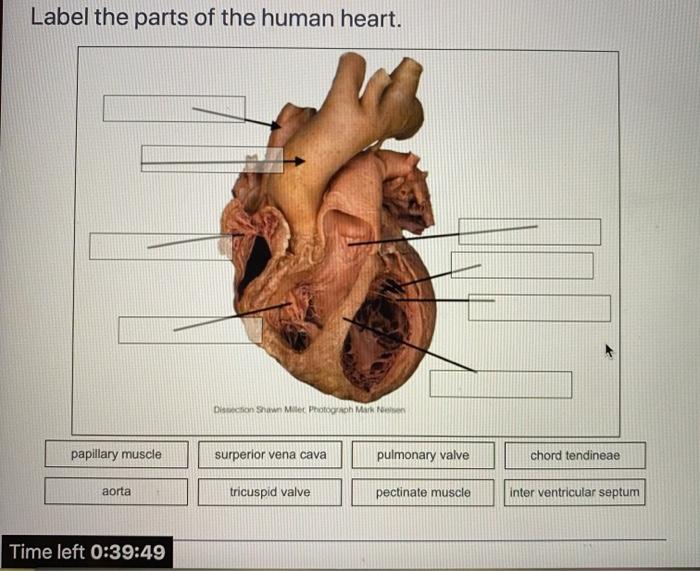
Solved Label the parts of the human heart. Dicson Shawn ...
Label parts of the heart - Liveworksheets Label parts of the heartDrag and drop the labels to the correct parts indicated on the heart diagram. ID: 832107. Language: English. School subject: Biology. Grade/level: GCSE. Age: 12-18. Main content: Label parts of the heart. Other contents: Add to my workbooks (517)
KS2 The Heart Diagram QR Labelling Activity (teacher made)
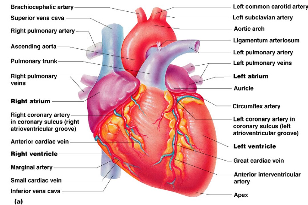
Heart anatomy: Structure, valves, coronary vessels | Kenhub Heart anatomy. The heart has five surfaces: base (posterior), diaphragmatic (inferior), sternocostal (anterior), and left and right pulmonary surfaces. It also has several margins: right, left, superior, and inferior: The right margin is the small section of the right atrium that extends between the superior and inferior vena cava .
Answered: Label the following parts of the heart… | bartleby
Human Heart - Diagram and Anatomy of the Heart - Innerbody The heart contains 4 chambers: the right atrium, left atrium, right ventricle, and left ventricle. The atria are smaller than the ventricles and have thinner, less muscular walls than the ventricles. The atria act as receiving chambers for blood, so they are connected to the veins that carry blood to the heart.
Heart Anatomy: Labeled Diagram, Structures, Blood Flow ...
Label the heart — Science Learning Hub Label the heart Interactive Add to collection In this interactive, you can label parts of the human heart. Drag and drop the text labels onto the boxes next to the diagram. Selecting or hovering over a box will highlight each area in the diagram. semilunar valve aorta left ventricle right ventricle left atrium vena cava pulmonary artery
Learn the Anatomy of the Heart
Heart: Anatomy and Function - Cleveland Clinic The parts of your heart are like the parts of a house. Your heart has: Walls. Chambers (rooms). Valves (doors). Blood vessels (plumbing). Electrical conduction system (electricity). Heart walls Your heart walls are the muscles that contract (squeeze) and relax to send blood throughout your body.

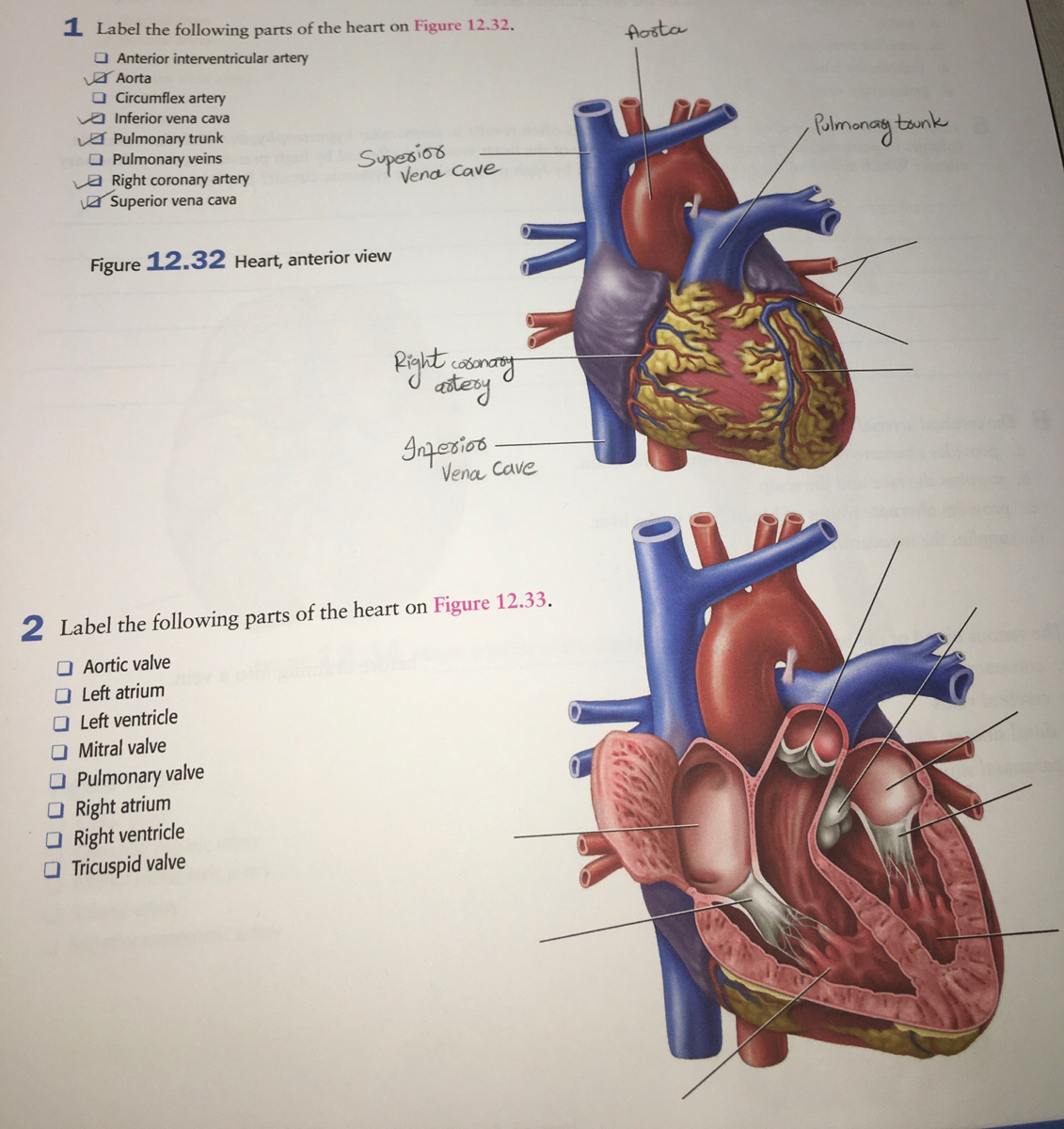
DIAGRAM OF THE HEART 1. Label the parts of the heart on the ...
Label the heart - Teaching resources
Human Heart Diagram Stock Photos, Pictures & Royalty-Free ...

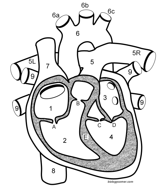
Label the parts 1to 9of the human heart in the figure given below
Heart | Structure, Function, Diagram, Anatomy, & Facts ...
Label the Heart Quiz

Heart Anatomy | Anatomy and Physiology II
Simple Heart Diagram with Labels Activity - Human Biology

pictures with parts labeled - Google Search | Human heart ...
Label the heart — Science Learning Hub
heartbeat2017-Online MPEG-4

File:Heart diagram-en.svg - Wikipedia

Label the Heart worksheet | KS2 | Primary Science | Distance ...

Draw a diagram of the human heart and label its parts ...

Pin on Paramedic Study Guide
Heart Anatomy/Heart Dissections/Heart Labeling ...
Parts of the Heart label & Diagram - Etsy

685 Human Heart 3d With Label Images, Stock Photos & Vectors ...
Real Heart Diagram Poster - Etsy

label the parts of human heart - Brainly.ph

Anatomy of the heart. A, Cross section of the heart wall ...

Human Anatomy: Label the Heart Worksheets in 3 Differentiated ...

II. label the parts of the heart - Brainly.ph
The Heart Diagram Label Worksheets (Differentiated ...

heart labeling Flashcards | Quizlet

Label the Heart's Parts Printables for 6th - 9th Grade ...

Human Heart Labeling Worksheet | Heart diagram, Heart ...
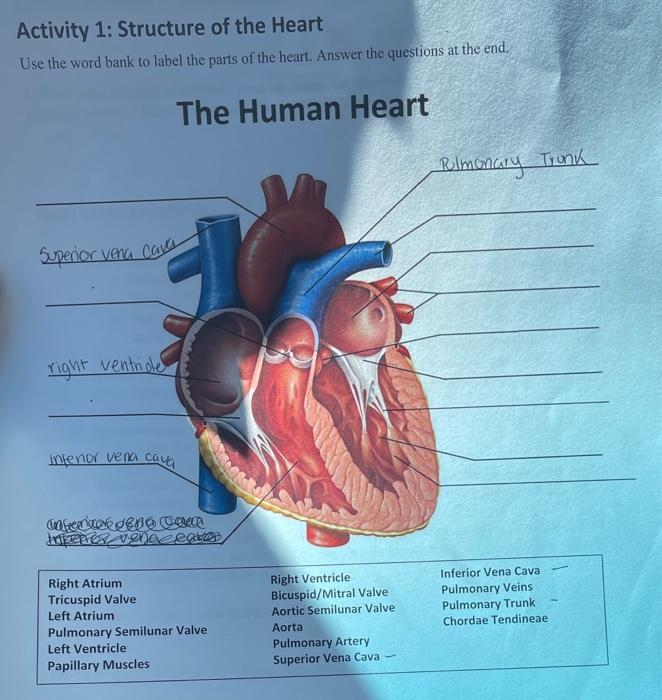
Solved Activity 1: Structure of the Heart Use the word bank ...

File:Diagram of the human heart (cropped).svg - Wikipedia
Draw a labelled diagram of the human heart and label its parts.
Parts Of The Heart - ProProfs Quiz

Draw a diagram of the sectional view of human seminiferous ...
Draw a diagram of the human heart and label its parts.
Draw the diagram showing the sectional view of the human ...

In a bond paper , you draw the human heart and label the ...
Label the heart — Science Learning Hub
Post a Comment for "43 label heart parts"