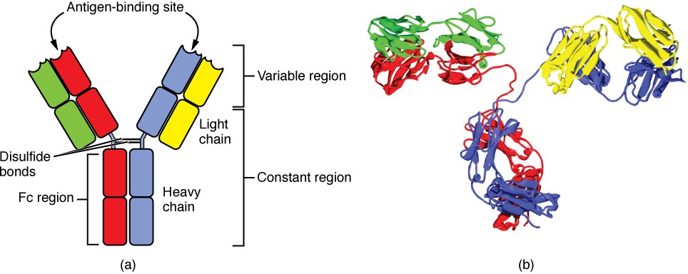
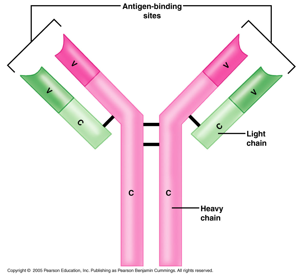
44 which label or labels indicate(s) the antigen binding site?
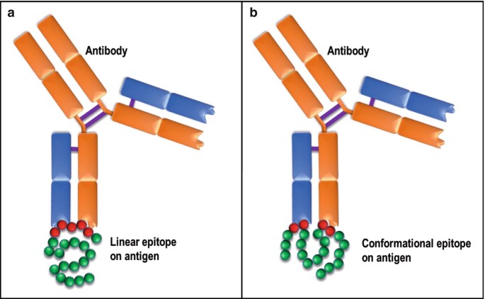
Frontiers | SEMA: Antigen B-cell conformational epitope prediction ... One of the primary tasks in vaccine design and development of immunotherapeutic drugs is to predict conformational B-cell epitopes corresponding to primary antibody binding sites within the antigen tertiary structure. To date, multiple approaches have been developed to address this issue. However, for a wide range of antigens their accuracy is limited. In this paper, we applied the transfer ... Unit Four: Lymphatic system and Immunity Flashcards | Quizlet In IgG, the antigen binding site is formed by. the variable segments of both the light and heavy chains. _____ is the class of antibody first secreted in response to a new antigen. IgM. ... Which label or labels indicate(s) the antigen binding site? The two sites labeled with the letter A.
Immunoassay Methods - Assay Guidance Manual - NCBI Bookshelf The critical steps in setting up a screen are as follows: 1. Develop a validated immunoassay as described above. 2. Acquire antibody, antigen/calibrator, label and buffer reagents in quantities needed for HTS. 3. Establish liquid handling and automation procedures for screening and immunoassay methods. 4.

Which label or labels indicate(s) the antigen binding site?
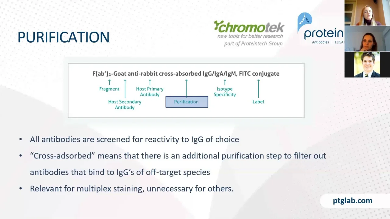
Immunolabeling | Thermo Fisher Scientific - US For direct immunofluorescence, the antibody binding the epitope is labeled with fluorophores (green). For indirect or secondary detection, the primary antibody binds the epitope and a fluorophore-labeled secondary antibody (purple) that has specificity for the primary antibody binds to it. Direct immunofluorescence Immunoassay - Wikipedia In addition to the binding of an antibody to its antigen, the other key feature of all immunoassays is a means to produce a measurable signal in response to the binding. ... Most, though not all, immunoassays involve chemically linking antibodies or antigens with some kind of detectable label. A large number of labels exist in modern ... Different Ways to Add Fluorescent Labels - Thermo Fisher Scientific Labeling various targets with separate fluorescent colors allows you to visualize different structures or proteins within a cell in the same experiment. Ways to fluorescently label your target include fluorescent dyes, immunolabeling, and fluorescent fusion proteins —all of which can provide a means to selectively mark structures and proteins ...
Which label or labels indicate(s) the antigen binding site?. Western Blot Technique: Principle, Steps, Uses | Microbe Online Sep 17, 2022 · This will prevent the non-specific binding of the antibody and reduce the overall background signal. Common blocking buffers include 5% non-fat dry milk or BSA in a TBS-Tween solution. However, do not use a milk solution when probing with phosphor-specific antibodies as it can cause high background from its endogenous phosphoprotein, casein. Guidelines for pre‐transfusion compatibility procedures in ... Final verification of the label and unit after attaching tags is recommended. Ideally, this should be an electronic check but could be a visual confirmation. 7.21.7. Blood component labels should only be printed and attached for one patient at a time to avoid the risk of transposition of labels between units for different patients. 7.21.8. Real-Time, label-free monitoring of tumor antigen and serum antibody ... The binding kinetics and affinity values of anti-NY-ESO-1 monoclonal antibody, ES121, to the cancer-testis antigen NY-ESO-1 were determined according to the surface heterogeneity model and resulted in K D values of 1.3×10 −9 and 2.1×10 −10 M. The reconfigured instrument was then used to measure the interaction between tumor antigens and ... Why Site-Specific — AlphaThera Site-specific antibody labeling ensures that the label does not interfere with antigen binding. This becomes particularly important when attaching large biomolecules to the antibody (e.g. enzymes), which can sterically prevent antigen binding. Site-specific labeling also ensures the proper orientation of immobilized antibodies.
dock8 deficiency attenuates microglia colonization in early ... Aug 17, 2022 · A Schematic diagram of the imaging region. Black dash lines represent the imaging area. B, E, H Representative images (B) and quantification of 4 dpf (E) and 6 dpf (H) NR signals of dock8-15,+5bp ... Leave for personal reasons (special leave) — Business ... Death of the worker's or the spouse's/partner's second-degree relative (grandparents, grandchildren, brothers and sisters, brothers-in-law and sisters-in-law) 1 day In the event of moving, the 2 days of special leave are granted only once over a 3-year period with the same employer unless the worker has to move for professional reasons. Anatomy Labs for Exam 2 Flashcards | Quizlet Which label or labels indicate(s) the antigen binding site? The two sites labeled with the letter A. What defense mechanism is shown in the images above? Complement (group of proteins that work together, binding to antibodies and forming large holes in target cells) What defense mechanism is shown in this image? Destruction by an NK cell. M A&P Introduction to Lymphatic System and Immunity Label the cause of infection and some structures involved in fighting the infection. After the proteins are separated by electrophoresis, the _____. ... Which label or labels indicate(s) the antigen binding site? the two sites labeled with the letter A. Steps in antigen presentation include which of these?
Learning context-aware structural representations to predict antigen ... our framework comprises three components to leverage biological insights: (i) graph convolutions to capture the spatial relationships of the interfaces, (ii) an attention layer to enable each protein's interface predictions to account for the potential binding context provided by its partner and (iii) transfer learning to leverage the larger set … EIAs and ELISAs | Microbiology | | Course Hero A red color (from gold particles) or blue (from latex beads) developing at the test line indicates a positive test. If the color only develops at the control line, the test is negative. Like ELISA techniques, lateral flow tests take advantage of antibody sandwiches, providing sensitivity and specificity. Antibody Types: IgM, IgA, IgD, IgG, IgE and Camelid Antibodies However, since pentameric IgM has 10 antigen binding sites, it has higher avidity (overall binding strength) for antigens than IgG and acts as an excellent activator of the complement system and ... Conformation of peptides bound to the transporter associated with ... For resolving the structure of bound peptides and the mechanism of peptide binding, we used EPR spectroscopy in combination with site-directed spin labeling of antigenic peptides to ( i) probe the spatial arrangement of the peptide-binding pocket of TAP and ( ii) to elucidate the conformation of the bound peptide. Go to: Results
Simple, Rapid Chemical Labeling and Screening of Antibodies ...
Exam 3: Mastering Lymphatic System (#6) Flashcards | Quizlet Which of these statements about lymphocytes is false? -They bind antigens. -They mostly occur in lymphoid tissues. -They are phagocytic. -They occur as B, T, and NK types. they are phagocytic Classes of lymphocytes T cells B cells NK cells T cells are approximately ___% of circulating lymphocytes. 80% B cells are ___% of circulating lymphocytes.

Site-Specific Antibody Labeling with oYo-Link
Structure and Function of Immunoglobulins - PMC - PubMed Central (PMC) The antigen binding site is the product of a nested gradient of diversity (A) H chain rearrangement can yield as many as 38,000 different VDJ combinations. The addition of nine N nucleotides on either side of the D gene segment yields can yield up to 64,000,000 different CDR-H3 junctional sequences.

A&P2 Lab 10 HW Flashcards | Quizlet
Part a drag the labels onto the diagram to identify - Course Hero HelpReset the two sites labeled with the letter A the single location labeled B the two sites labeled C the two sites labeled D Heavy chain Antigen biding site Variable segment Light chain Constant segments of light and heavy chains Complement binding site Site of binding to macrophages

Chapter 1, 5, 10 and 14-17 Study Questions Flashcards | Quizlet
antigens are found naturally on a particle Examples of direct ... Capture antibody is first attached to a solid phase 2. Unknown patient antigen (usually serum) is then allowed to react with the antibody 3. After washing to remove unbound antigen, a second antibody with a label is added to the reaction How is a labeled analyte detected? By measuring radioactivity in RIA molecule (radioimmunoassay)

A&P2 Lab 13 HW, A&P2 Lab 12 HW, A&P2 Lab 11 HW, A&P2 Lab 10 ...
Structure of Antibody (With Diagram) | Organisms - Biology This variable region, composed of 110-130 amino acids, gives the antibody its specificity for binding antigen. ADVERTISEMENTS: Antibodies are of five classes - IgG, IgA, IgM, IgD and IgE. Ig stands for immunoglobulins. IgG constitutes to about 75% of the total antibodies. IgE is involved in allergy and IgM is formed during the primary response.
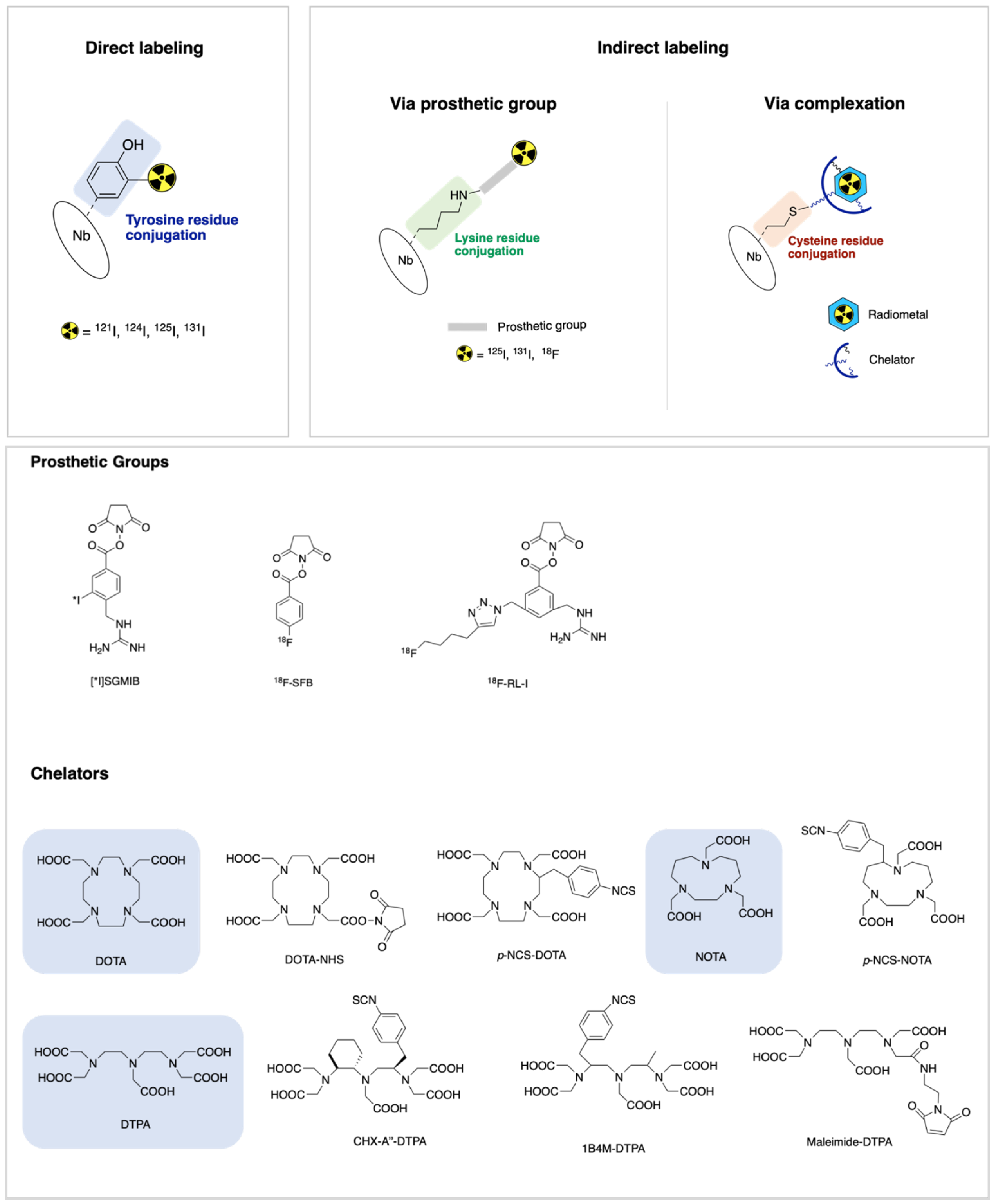
IJMS | Free Full-Text | Nanobody-Based Theranostic Agents for ...
Multiplex label-free biosensor for detection of ... - ScienceDirect The labels may destroy the binding sites that participate in antibody-antigen interactions, on the one hand, and non-specifically interact with proteins immobilized on a surface, on the other hand (Cooper, 2009; Fan et al., 2008; Orlov et al., 2018; Seydack, 2005). The label-free techniques are not prone to the label-associated issues.

Draw a neat labeled diagram of an antibody molecule and ...
Antibody Structure - University of Arizona An antigenic determinant, a site on the antigen that the immune system responds to by making antibody, can frequently be one unique structure on the antigen. In hen egg white lysozyme, a glutamine at position 121 (Gln 121) protrudes away from the antigen surface. In this view, Gln 121 is circled. The antibody is not shown.
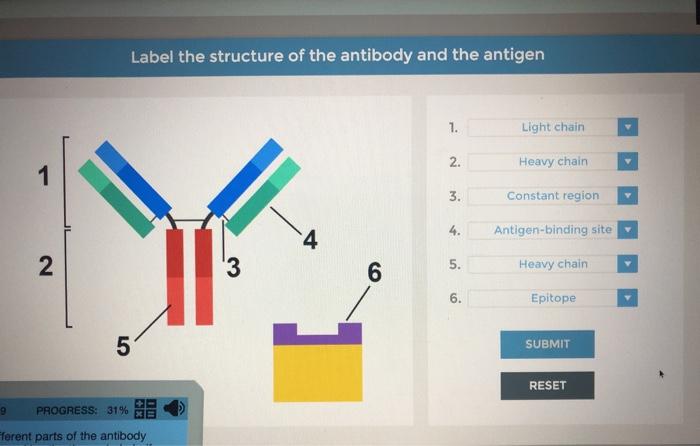
Solved Label the structure of the antibody and the antigen ...
DailyMed - LUPRON DEPOT- leuprolide acetate kit In vitro binding to human plasma proteins ranged from 43% to 49%. Elimination The mean systemic clearance of leuprolide following intravenous bolus administration to healthy male volunteers was 7.6 L/h, and terminal elimination half-life was approximately 3 hours based on a two compartment model.
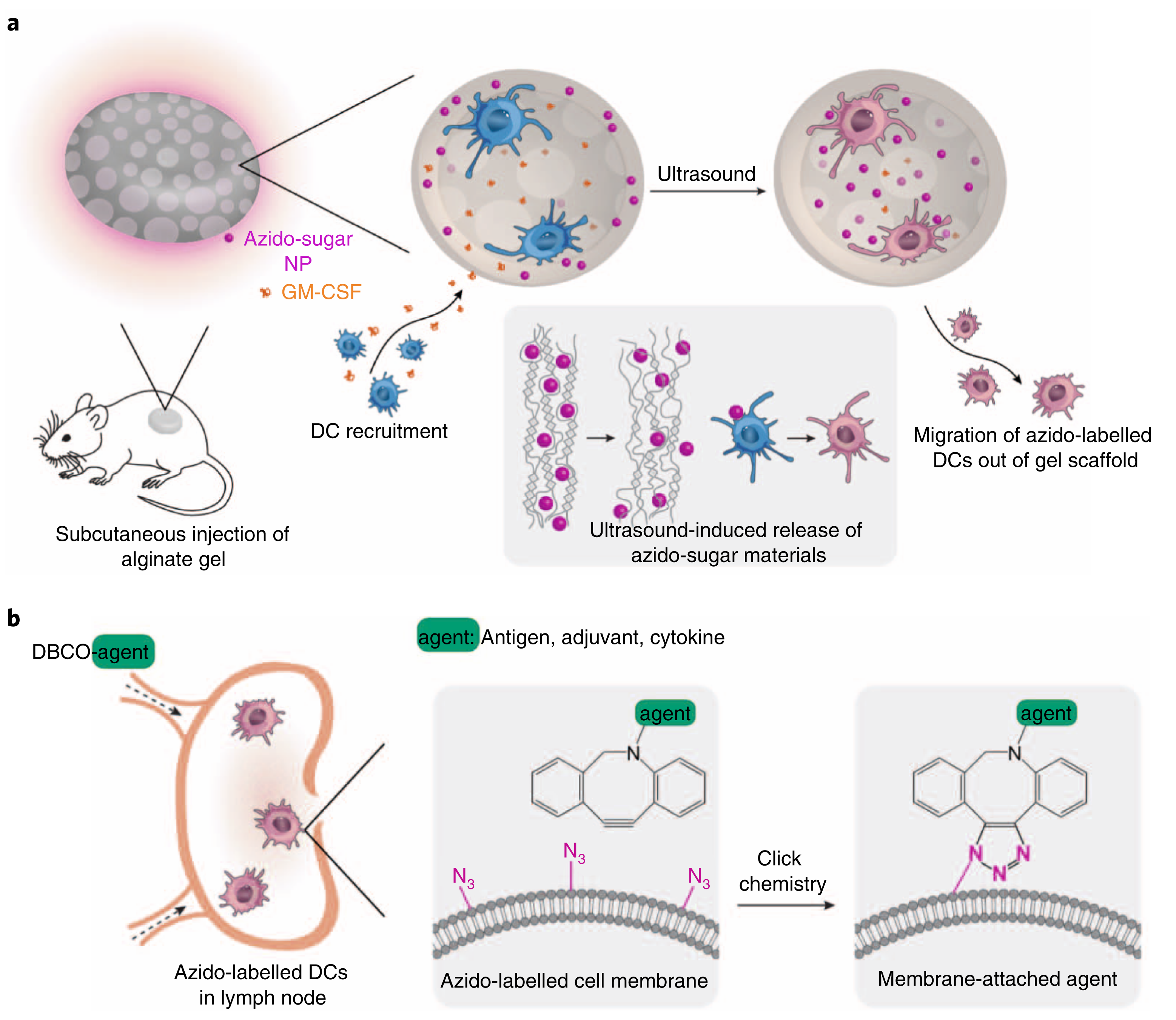
Metabolic labeling and targeted modulation of dendritic cells ...
Antibody Conjugation One method to reduce the number of undesirable conjugations is the replacement of cysteine with selenocysteine. This reduces the probability of conjugation occurring at inappropriate sites of the polypeptide chain(s) that disrupt the structure of the antibody (and therefore its affinity and/or specificity) [].In the case of antibody-drug conjugates, modification of glutamine residues using the ...
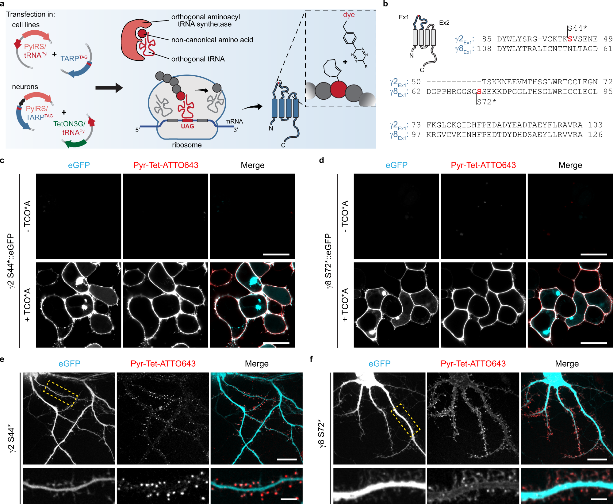
Bioorthogonal labeling of transmembrane proteins with non ...
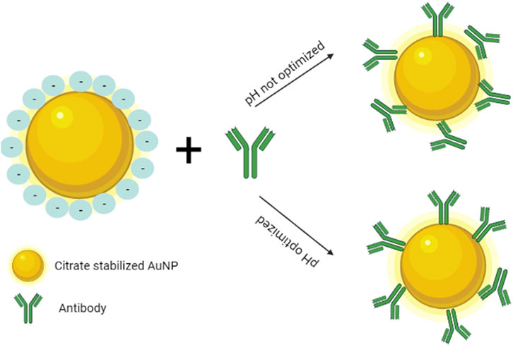
Lateral flow assays: Principles, designs and labels An alternative biorecognition molecule of antibody is immunogen, which is a cheaper biomolecule than an antibody, since the production cost of antibody is high. The binding of antibody to colloidal gold occurs by means of hydrophobic residues in the antigen-binding site of antibody. This way, the binding sites of an antibody are decreased.

Ch. 16- Prep for Exam Flashcards | Quizlet
US5601986A - Assays and devices for the detection of ... The present invention involves a variety of assay methods and devices for screening or diagnosing the occurrence of extrahepatic biliary atresia. In particular the methods and devices involve an antibody specifically for the detection of dipeptidyl peptidase IV in a test sample as indicative of extrahepatic biliary atresia.
Chapter 8: THE LYMPHATIC AND IMMUNE SYSTEM – Anatomy & Physiology
PDF Secondary - Novus Bio One antigen binding site Reduce non-specific binding between Fc receptors and the Fc portion of the antibody. Fragment antibodies penetrate tissue better than whole antibodies due to their smaller size. This may enhance staining in IHC. F(ab') 2 Two arms of the antibody Divalent: Two antigen binding sites Reduce non-specific binding

Chemical Profiling of the Endoplasmic Reticulum Proteome ...
HW 29.docx - Best of Home Work (Exercise 29: Blood) Drag... Label the red blood cells with the correct antigen (s). Left to right… a antigen, b antigen, a & b, none Left to right … a antigen , b antigen , a & b , none Antibodies are proteins that have a lock-and-key recognition for their antigen established by the antigen-binding site on the antibody.

GFP nanobodies enable the selective labeling of newly ...
Abzymes: Catalytic Antibodies - UC Santa Barbara Rotate around active site complex. Label charged or partially charged centers. Labels off. For best results, push the buttons in sequence. 1. Zoom into the antigen-binding site with bound hapten. Initially, the ... The germline sequences of the heavy chain gene segments indicate that this particular asparagine residue arose as the result of ...

Secondary Antibodies and Conjugates - PDF Free Download
Exam 2 Lab Flashcards | Chegg.com Which label or labels indicate (s) the antigen binding sight? The two sides labeled D The two sides labeled A The single location labeled B The two sites labeled C A What defense mechanism is shown in the images above? Complement Pyrogen Interferon Perforin Complement What defense mechanism is shown in this image?

An example monoclonal antibody structure (pdb 1IGT, mouse ...
A&P2 Lab 10 HW Flashcards | Quizlet Study with Quizlet and memorize flashcards containing terms like Drag the labels onto the diagram to identify the lymphoid tissues and organs of the lymphatic system., Drag the labels onto the diagram to identify the structural features of the spleen., Which of the labels indicates a structure through which lymph flows? and more.
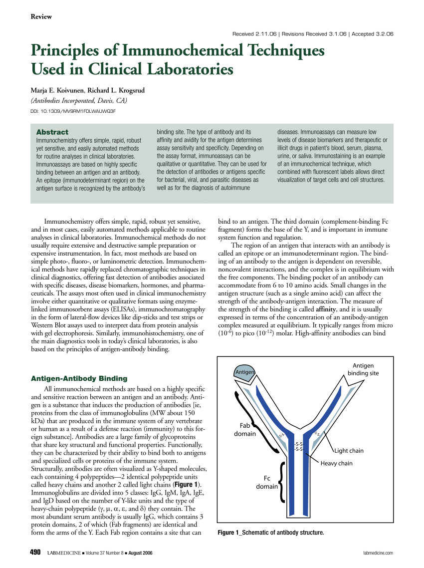
PDF) Principles of Immunochemical Techniques Used in Clinical ...
Different Ways to Add Fluorescent Labels - Thermo Fisher Scientific Labeling various targets with separate fluorescent colors allows you to visualize different structures or proteins within a cell in the same experiment. Ways to fluorescently label your target include fluorescent dyes, immunolabeling, and fluorescent fusion proteins —all of which can provide a means to selectively mark structures and proteins ...
Frontiers | Latest Trends in Lateral Flow Immunoassay (LFIA ...
Immunoassay - Wikipedia In addition to the binding of an antibody to its antigen, the other key feature of all immunoassays is a means to produce a measurable signal in response to the binding. ... Most, though not all, immunoassays involve chemically linking antibodies or antigens with some kind of detectable label. A large number of labels exist in modern ...

A&P2 Lab 10 HW Flashcards | Quizlet
Immunolabeling | Thermo Fisher Scientific - US For direct immunofluorescence, the antibody binding the epitope is labeled with fluorophores (green). For indirect or secondary detection, the primary antibody binds the epitope and a fluorophore-labeled secondary antibody (purple) that has specificity for the primary antibody binds to it. Direct immunofluorescence
Immune System

Affinity-Based Methods for Site-Specific Conjugation of ...
Selecting the right antibodies and nanobodies for Fluorescent Imaging
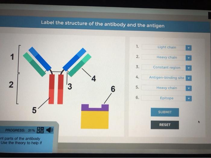
Solved Label the structure of the antibody and the antigen ...

Electron Microscopic Detection of Single Membrane Proteins by ...

A&P2 Lab 10 HW Flashcards | Quizlet

Multicolor labeling of HA-tagged proteins in vivo. a ...

Analysis of Cell-Surface Receptor Dynamics through Covalent ...

Systematic identification of engineered methionines and ...
Immunoassays | SpringerLink

Antibody Structure

Determining Binding Affinity (KD) of Radiolabeled Antibodies ...

A&P2 Lab 13 HW, A&P2 Lab 12 HW, A&P2 Lab 11 HW, A&P2 Lab 10 ...
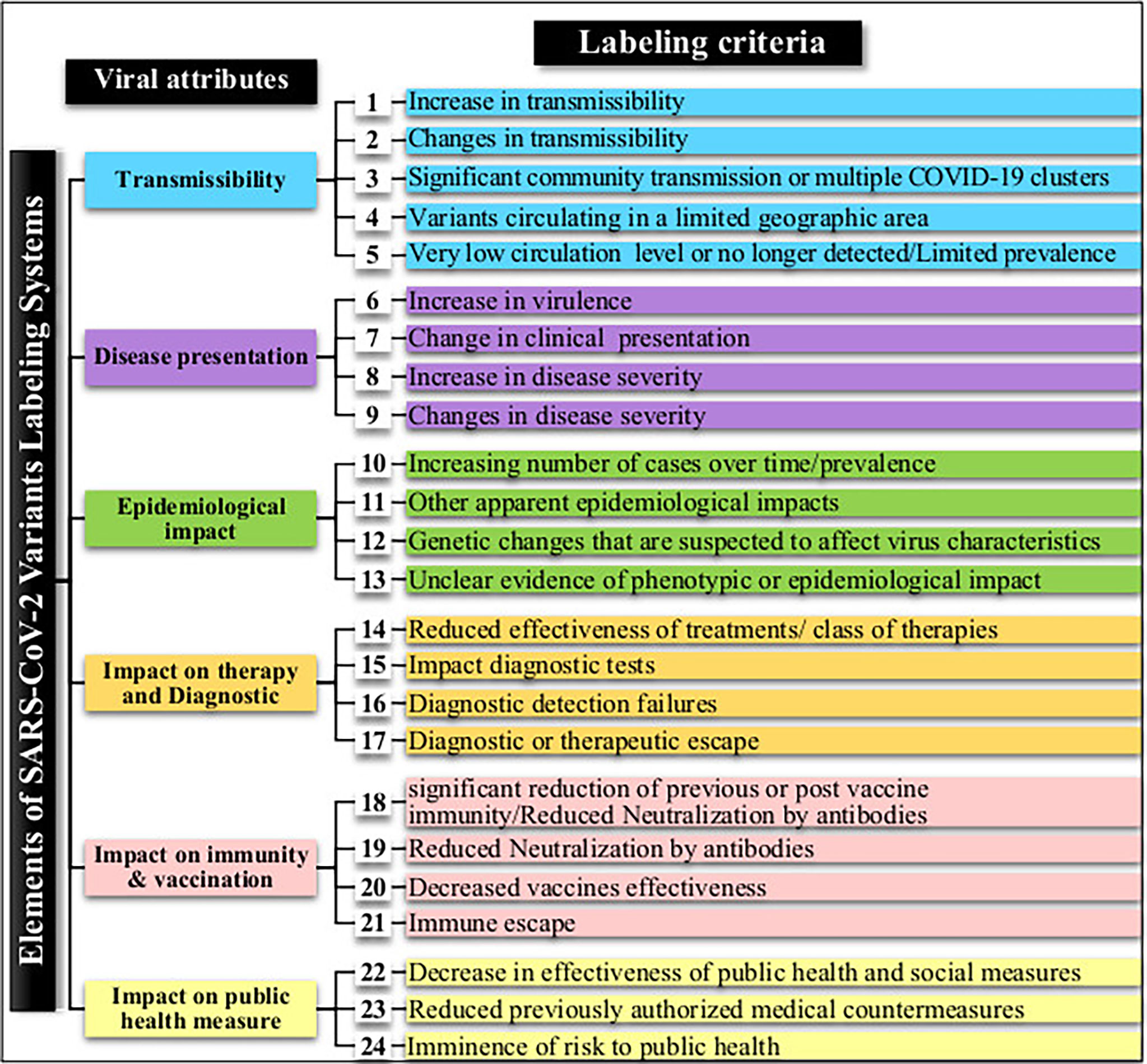
Frontiers | How concerning is a SARS-CoV-2 variant of concern ...

Analysis of Cell-Surface Receptor Dynamics through Covalent ...
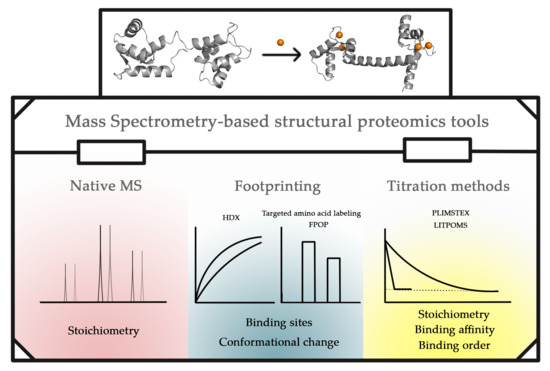
Biomolecules | Free Full-Text | Mass Spectrometry-Based ...
New method for localizing proteins in periodic structures ...

PDF) Multi-target immunofluorescence by separation of ...

Phenotypic determinism and stochasticity in antibody ...

PDF) Signal Enhancement in Oriented Immunosorbent Assays: A ...

Ch. 16- Prep for Exam Flashcards | Quizlet

Benchmarking UbiFast a Experimental design for TMT10-based ...

PDF) Multi-target immunofluorescence by separation of ...

Introduction of an Aldehyde Handle on Nanobodies by Affinity ...
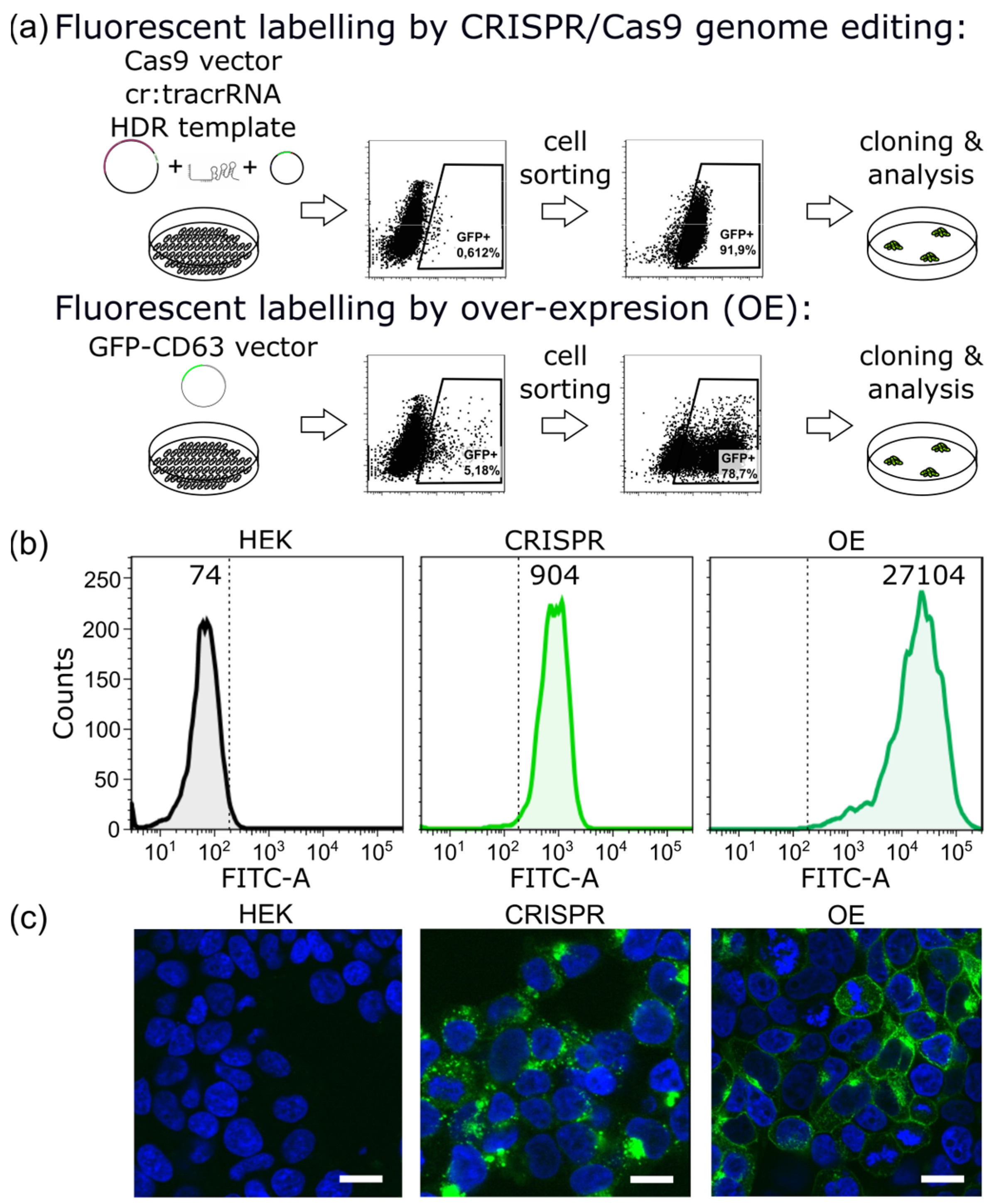
IJMS | Free Full-Text | CRISPR/Cas9 Genome Editing vs. Over ...
Post a Comment for "44 which label or labels indicate(s) the antigen binding site?"