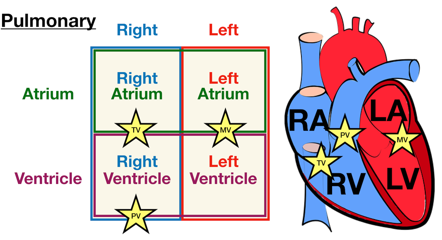
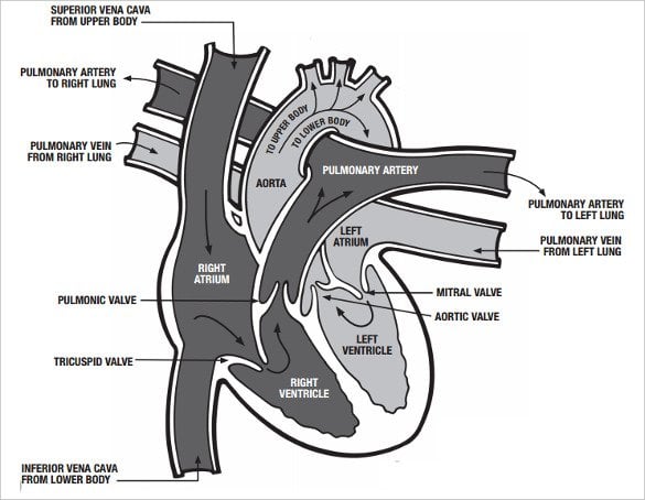
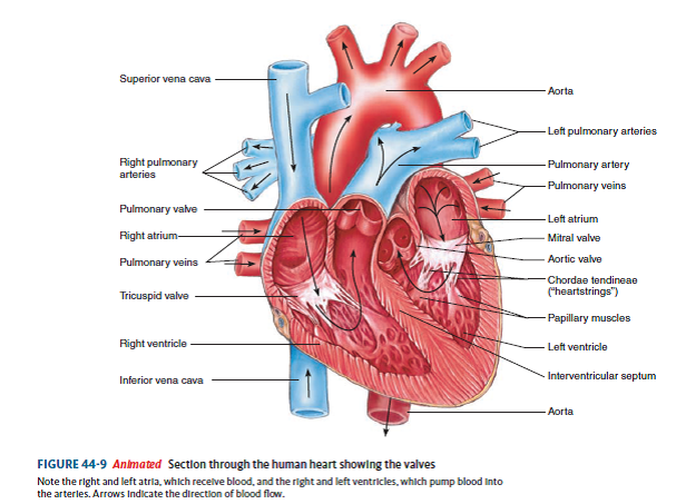
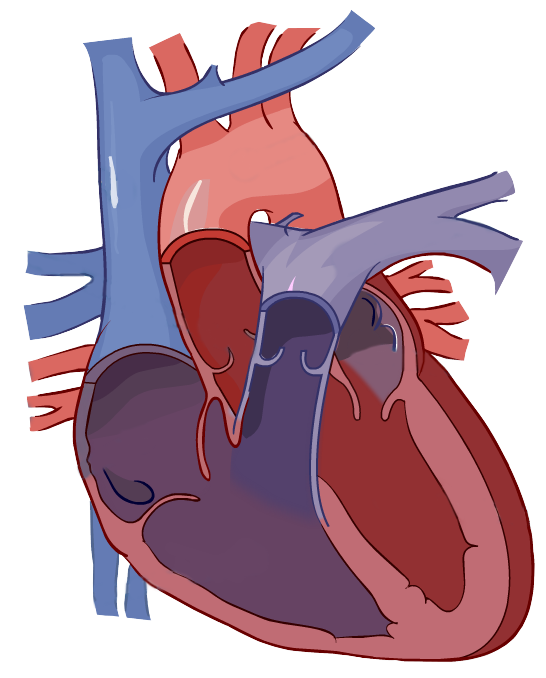
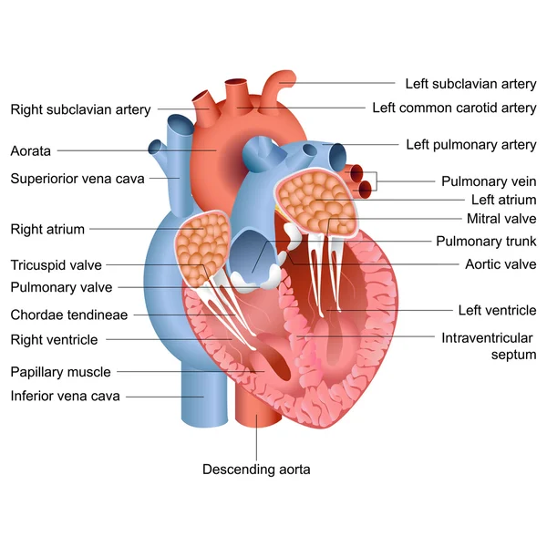
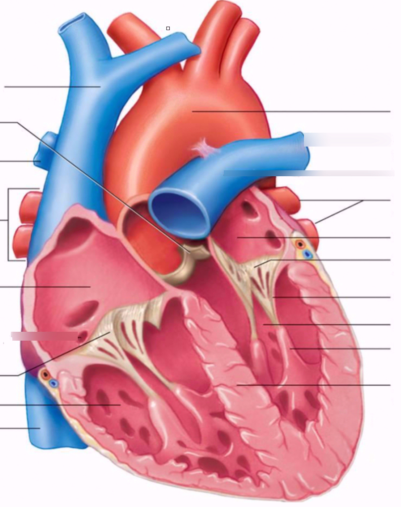
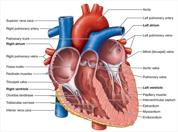
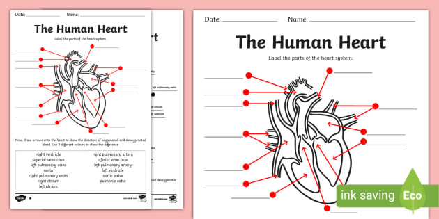
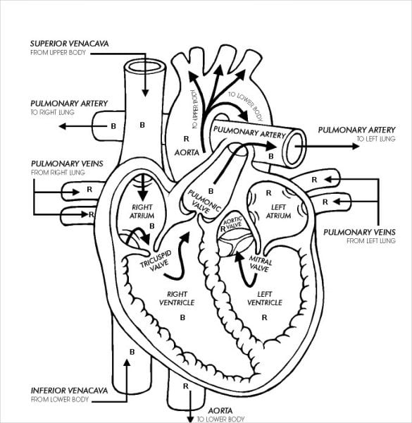
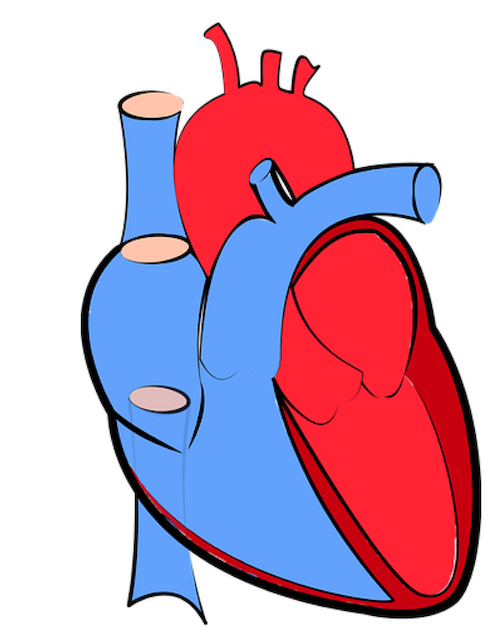
40 heart diagram labelling
Brigitte Zimmer Human Heart Diagram And Label. 9Th Grade Easy Plant Cell Diagram. Simple Diagram Of A Heart With Labels. Label The Picture Of A Human Digestive System. Labeled Sketch Of A Chloroplast. Internal Female Reproductive System Labelled Diagram. Dna Easy Drawing. Digestive System Diagram Class 10 Easy. Easy Plant Cell And Animal Cell Drawing With Labels. Charts of Normal Resting and Exercising Heart Rate Normal Heart Rate Chart During Exercise. Your maximum heart rate is the highest heart rate that is achieved during strenuous exercise. One method to calculate your approximate maximum heart rate is the formula: 220 - (your age) = approximate maximum heart rate. For example, a 30 year old's approximate maximum heart rate is 220 - 30 = 190 beats/min.
Conduction system of the heart: Parts and Functions - Kenhub The parts of the heart conduction system can be divided into those that generate action potentials (nodal tissue) and those that conduct them (conducting fibers). Although all parts have the ability to generate action potentials and thus heart contractions, the sinuatrial (SA) node is the primary impulse initiator and regulator in a healthy heart.

Heart diagram labelling
Origin and function of activated fibroblast states during zebrafish ... The adult zebrafish heart has a high capacity for regeneration following injury. ... We generated and characterized a transgenic line labelling col12a1a ... Venn diagram of upregulated genes in ... Alveoli: Function, Structure, and Lung Disorders - Verywell Health Alveoli are tiny, balloon-shaped air sacs in your lungs. The function of the alveoli is to move oxygen and carbon dioxide (CO2) molecules into and out of your bloodstream. Alveoli are an important part of your respiratory system, which includes the parts of your body that help you breathe . This article discusses the structure and function of ... Label a Nephron Quiz - PurposeGames.com This is an online quiz called Label a Nephron. There is a printable worksheet available for download here so you can take the quiz with pen and paper. From the quiz author. This is a nephron. Label it. This quiz has tags. Click on the tags below to find other quizzes on the same subject. biology. kidney. Nephron.
Heart diagram labelling. Circulatory System Diagram - New Health Advisor This circuit typically includes the movement of blood inside heart and 'myocardium' (the membrane of heart). Coronary circuit mainly consists of cardiac veins including anterior cardiac vein, small vein, middle vein and great (large) cardiac vein. There are different types of circulatory system diagrams; some have labels while others don't. Carpal bone quizzes and labeled diagrams | Kenhub Carpal bones labeled and unlabeled. The carpal bones names are labeled on the first diagram, while the second diagram is completely blank, ready for you to fill in for yourself. Don't worry if you don't get everything right the first time around - it's easy to get these carpal bones names mixed up, or for them to fly out of your memory ... How to Read an ECG | ECG Interpretation | EKG | Geeky Medics Confirm details. Before beginning ECG interpretation, you should check the following details: Confirm the name and date of birth of the patient matches the details on the ECG. Check the date and time that the ECG was performed. Check the calibration of the ECG (usually 25mm/s and 10mm/1mV). Mechanics of Breathing - Inspiration - TeachMePhysiology The Lungs and Breathing. The space between the outer surface of the lungs and inner thoracic wall is known as the pleural space.This is usually filled with pleural fluid, forming a seal which holds the lungs against the thoracic wall by the force of surface tension.This seal ensures that when the thoracic cavity expands or reduces, the lungs undergo expansion or reduction in size accordingly.
Root Cause Analysis: Definition and Examples | SafetyCulture The Root Cause Analysis Process in 5 Simple Steps. Root Cause Analysis Process. As shown in the diagram above, the root cause analysis steps are: Realize the problem. Gather data. Determine possible causal factors. Identify the root cause. Recommend and implement solutions. Going through each step in detail, here's how you can perform root ... Path of Blood Through the Heart - New Health Advisor Basics Parts of the Heart. Understanding the function of the heart is helpful to learn more about its anatomy. Here are the basic parts of the heart: 1. Right Atrium. The heart can be divided into right and left halves, as well as into the upper and lower chambers. There are two upper chambers called atria and two lower chambers called ventricles. disetar curriculum Read more label heart anatomy Labeling circulatory liveworksheets labelling igcse. July 14, ... If you are searching about Internal Organs Diagram - Online Dictionary for Kids… Read more Location of Heart in Human Body Hyaline cartilage - human body help. July 13, 2022 Post a Comment Understanding an ECG | ECG Interpretation | Geeky Medics An ECG electrode is a conductive pad that is attached to the skin to record electrical activity. An ECG lead is a graphical representation of the heart's electrical activity which is calculated by analysing data from several ECG electrodes. A 12-lead ECG records 12 leads, producing 12 separate graphs on a piece of ECG paper.
Penis: Anatomy, Function, Disorders, and Diagnosis Anatomy. The penis is one of the external organs of the male reproductive system (used for sex and to conceive babies) and the urinary system (used to "pee"). It is located at the front of the body at the base of the pelvis. The scrotum, containing the testes (a.k.a. testicles), is situated just beneath the penis. Double Circulation of Blood: Definition, Diagram - Embibe Fig: Double Circulation. Learn 12th CBSE Exam Concepts. Components Involved in Double Circulation. 1. Heart- It is four-chambered in humans, i.e., right and left atrium & right and left ventricle. 2. Blood vessels- Arteries, veins and capillaries come under blood vessels.Arteries carry oxygenated blood; veins carry deoxygenated blood except for the pulmonary artery and pulmonary vein. WHMIS 2015 - Labels : OSH Answers Suppliers and employers must use and follow the WHMIS 2015 requirements for labels and safety data sheets (SDSs) for hazardous products sold, distributed, or imported into Canada. Please refer to the following other OSH Answers documents for more information: WHMIS 2015 - General. WHMIS 2015 - Pictograms. Heart - Wikipedia The human heart is situated in the mediastinum, at the level of thoracic vertebrae T5-T8.A double-membraned sac called the pericardium surrounds the heart and attaches to the mediastinum. The back surface of the heart lies near the vertebral column, and the front surface sits behind the sternum and rib cartilages. The upper part of the heart is the attachment point for several large blood ...
Important Question for Class 10 Science Life Processes - Learn CBSE Draw a diagram of human excretory system and label kidneys, ureters on it. (Board Term I, 2017) Answer: Diagram of human excretory system is as follows: Question 37. Draw a neat diagram of excretory system of human beings and label on it: (i) Left kidney (ii) Urinary bladder. (Board Term I, 2016) Answer: Refer to answer 36. Question 38.
Know Where Your Heart Is and How to Identify Heart Pain Here we are going to discuss the symptoms of several chest pains which are associated with heart. 1. Heart Attack. Heart attack results from the occluded blood vessels that carry blood to the heart. The patient may experience the following signs: Fullness or squeezing sensation in the chest.
Female Body Diagram - Verywell Health Vagina: The vagina is a muscular canal that connects the cervix and the uterus, leading to the outside of the body. Parts of the vagina are rich in collagen and elastin, which give it the ability to expand during sexual stimulation and childbirth. Cervix: The cervix is the lower part of the uterus that separates the lower uterus and the vagina and may play a role in lubrication.
Layers of the heart: Epicardium, myocardium, endocardium - Kenhub The myocardium is functionally the main constituent of the heart and the thickest layer of all three heart layers. It is a muscle layer that enables heart contractions. Histologically, the myocardium is comprised of cardiomyocytes.Cardiomyocytes have a single nucleus in the center of the cell, which helps to distinguish them from skeletal muscle cells that have multiple nuclei dispersed in the ...
Labeled Periodic Table of Elements with Name [PDF & PNG] There are 118 elements in the periodic table, out of which 94 elements are natural, and others are nuclear reactor or laboratory tested elements. There are 18 groups in the periodic table, which consists of metal and nonmetal. Protons in the tables are positively charged particles. Neutrons are the neutrally negative charge, and electrons are ...
Know the Structures and Functions about Your Heart The human heart is just roughly about the size of a fist. Your heart weighs about 10 - 12 ounces (or 280 - 340 grams) if you are a man, and 8 -10 ounces (or 230 - 280 grams) if you are a woman. In an adult, the heart beats at an average of 60-80 times per minute. The newborn's heart beats faster than an adult heart, at about 70-190 beats/minute.
Heart valves anatomy: Tricuspid-aortic-mitral-pulmonary - Kenhub Right atrioventricular valve. Valva atrioventricularis dextra. 1/4. Synonyms: Tricuspid valve, Valva tricuspidalis. Understanding heart valves anatomy is important in grasping the overall function of the heart. The heart is one of the most important organs in the body. It is responsible for propelling blood to every organ system, including itself.
Of The Heart Worksheet Anatomy Anatomy of the Heart Gross Anatomy of the Human Heart 1 The heart is placed obliquely behind the body of the sternum and adjoining parts of the costal cartilages so that one-third of it lies to the right and two-thirds to the left of the median plane The Heart, part 2 - Heart Throbs: Crash Course A&P #26 The frog heart, however, has only one lower chamber, a single ventricle Outside View of ...
Circulatory system - Wikipedia The circulatory system includes the heart, blood vessels, and blood. The cardiovascular system in all vertebrates, consists of the heart and blood vessels. The circulatory system is further divided into two major circuits - a pulmonary circulation, and a systemic circulation. The pulmonary circulation is a circuit loop from the right heart taking deoxygenated blood to the lungs where it is ...
DP Biology: Heart Dissection & Diagram - ThinkIB Heart Dissection & Diagram. Using a short screen cast students learn a simple method of drawing a diagram of the heart. Annotations are added and students can practice their knowledge using some printable flashcards or a range of online study methods. Students then extend their knowledge by completing a detailed heart dissection.
Heart anatomy: Structure, valves, coronary vessels - Kenhub Heart anatomy. The heart has five surfaces: base (posterior), diaphragmatic (inferior), sternocostal (anterior), and left and right pulmonary surfaces. It also has several margins: right, left, superior, and inferior: The right margin is the small section of the right atrium that extends between the superior and inferior vena cava .
Label a Nephron Quiz - PurposeGames.com This is an online quiz called Label a Nephron. There is a printable worksheet available for download here so you can take the quiz with pen and paper. From the quiz author. This is a nephron. Label it. This quiz has tags. Click on the tags below to find other quizzes on the same subject. biology. kidney. Nephron.
Alveoli: Function, Structure, and Lung Disorders - Verywell Health Alveoli are tiny, balloon-shaped air sacs in your lungs. The function of the alveoli is to move oxygen and carbon dioxide (CO2) molecules into and out of your bloodstream. Alveoli are an important part of your respiratory system, which includes the parts of your body that help you breathe . This article discusses the structure and function of ...
Origin and function of activated fibroblast states during zebrafish ... The adult zebrafish heart has a high capacity for regeneration following injury. ... We generated and characterized a transgenic line labelling col12a1a ... Venn diagram of upregulated genes in ...












Post a Comment for "40 heart diagram labelling"