40 sarcomere diagram labeled
Titin - Wikipedia Titin / ˈ t aɪ t ɪ n / (contraction for Titan protein) (also called connectin) is a protein that in humans is encoded by the TTN gene. Titin is a giant protein, greater than 1 µm in length, that functions as a molecular spring which is responsible for the passive elasticity of muscle.It comprises 244 individually folded protein domains connected by unstructured peptide sequences. Sarcomere | Cell and Developmental Biology | SUNY Upstate Medical ... Sarcomere. Diagram of a sarcomere bounded by the Z-bands. The left side (peach color) of the sarcomere represents a half sarcomere found in vertebrate skeletal myofibrils. Note that the nebulin molecules are part of and extend the entrie length of the thin filaments. The right side (pink color) of the sarcomere reflects a half sarcomere in ...
Sarcomere - an overview | ScienceDirect Topics A sarcomere is the basic contractile unit of muscle fiber. Each sarcomere is composed of two main protein filaments—actin and myosin—which are the active structures responsible for muscular contraction. ... Figure 2-6 diagrams the cytoskeleton of the sarcomere and its relationship to the contractile proteins. 6 The M line and the Z disc ...

Sarcomere diagram labeled
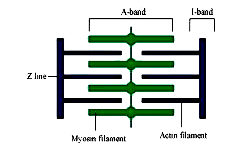
Home Page: Journal of Hand Surgery 26.05.2022 · The Journal of Hand Surgery publishes original, peer-reviewed articles related to the pathophysiology, diagnosis, and treatment of diseases and conditions of the upper extremity; these include both clinical and basic science studies, along with case reports.Special features include Review Articles (including Current Concepts and The Hand Surgery Landscape), … Structure and Function of the Skeletal Muscle Extracellular Matrix 01.09.2011 · Furthermore, multiple reaction monitoring may be used in the future to quantify proteins from labeled cell types in muscle. 107 This method can detect and quantify peptides in multiplex format using a triple quadrupole instrument. An advantage to this method is that it might be used to identify protein signals in fibrotic muscle that cause cells to overexpress collagen. Question Video: Identifying the Z Line in the Sarcomere | Nagwa Two successive Z lines mark the boundary of each sarcomere. The A bands, labeled here with the letter Z, are the regions of the sarcomere that do contain the thicker, darker myosin filaments. And so they are sometimes called the dark bands. The outer edges of the A bands are darkest as they contain both actin and myosin filaments overlapping.
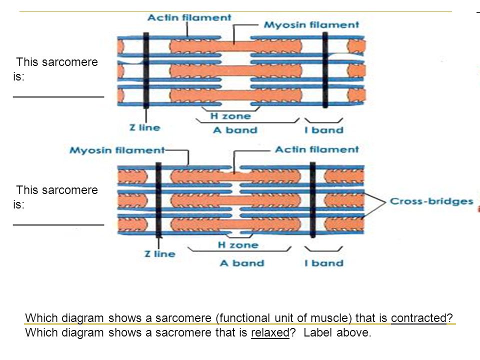
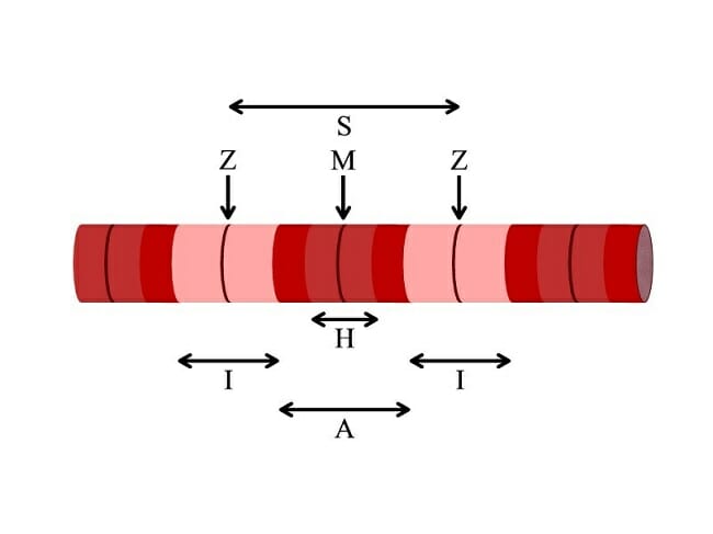
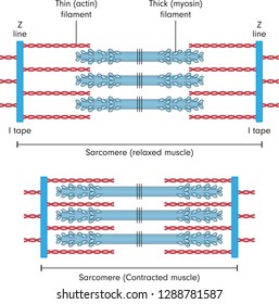
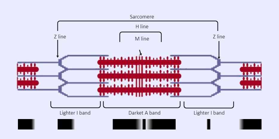
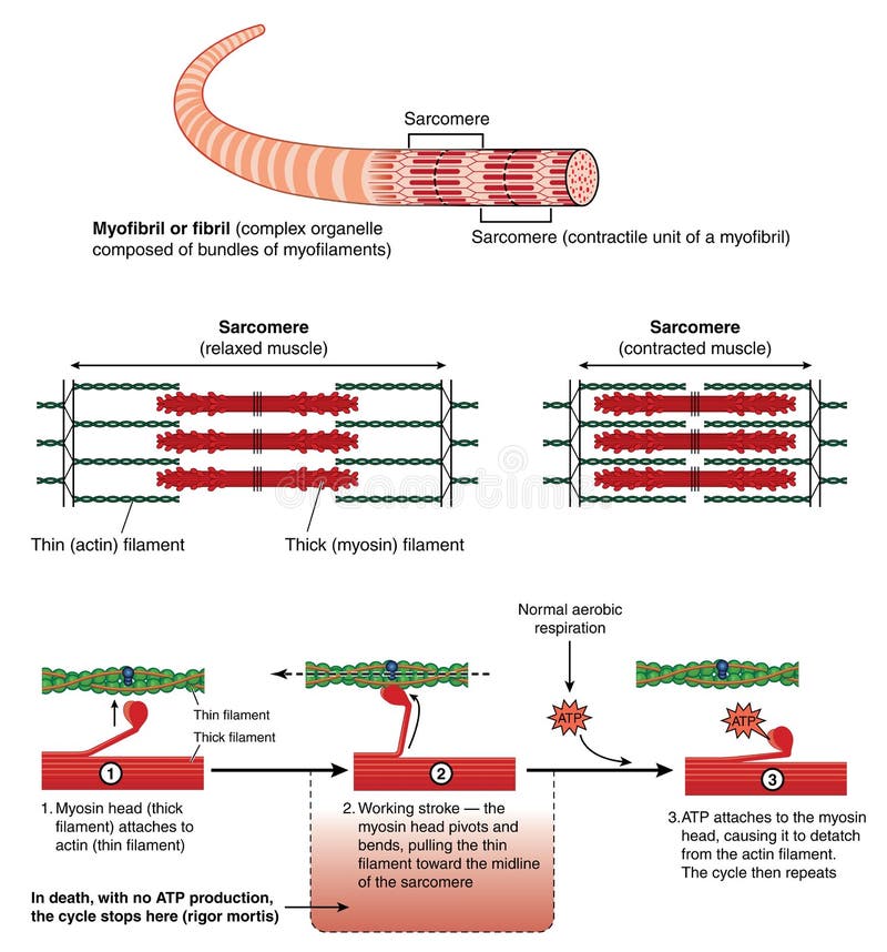
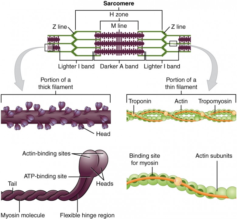
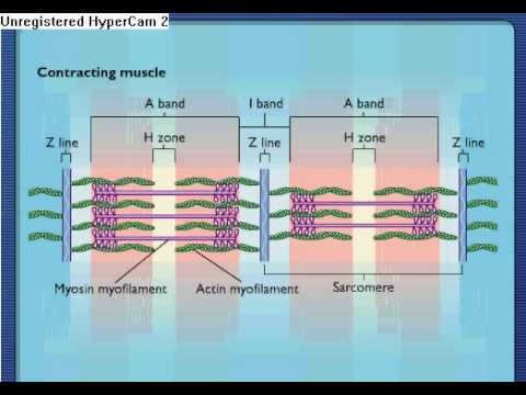
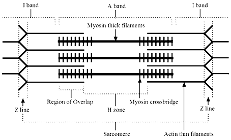
Sarcomere diagram labeled. Sarcomeres: "I" and "A" Bands, "M" and "Z" Lines, "H" Zone • A sarcomere is the basic contractile unit of skeletal muscle that is made of thick and thin filaments. • Thick filaments are organized bundles of myosin, while thin filaments are made of actin along with the two other regulatory proteins-troponin and tropomyosin. • Z-lines define the boundaries of each sarcomere. McGraw Hill Legacy Resources | Glencoe, SRA, and Macmillan Health (6–12) Teen Health and Glencoe Health are application-based programs that teach the 10 critical health skills that align with the National Health Standards. While emphasizing social and emotional skills, these programs explore up-to-date information and statistics on timely, relevant topics to help students become health-literate individuals. Sarcomere- Definition, Structure, Diagram, and Functions A sarcomere is a complex multicomponent biological system and functional unit of striated muscle which plays a vital role in transforming the chemical energy released upon the ATP hydrolysis into mechanical work. Skeletal muscles are made up of the basic unit called a sarcomere and all voluntary movement is initiated by this skeletal muscle. sarcomere Diagram | Quizlet Sarcomere. contractile unit of a muscle fiber. what area of the sarcomere will shorten during muscle contraction. i band and H zone will shorten during contraction. what is actin? This term refers to a thin protein filament that acts with myosin filaments to produce muscle action.
Sarcomere Labeling Diagram | Quizlet Sarcomere Labeling Diagram | Quizlet Textbook solutions Sarcomere Labeling STUDY Learn Write Test PLAY Match + − Created by Colby_Felton-Bettis Terms in this set (7) Sarcomere ... H zone ... M line ... Actin ... Z line ... A band ... I band ... Sets found in the same folder Synapses Labeling 8 terms Colby_Felton-Bettis Ear Labeling 8 terms Sarcomere Structure Labeled - welcome to mozhgon jeddi joins the lab ... Sarcomere Structure Labeled. Here are a number of highest rated Sarcomere Structure Labeled pictures upon internet. We identified it from reliable source. Its submitted by executive in the best field. We assume this kind of Sarcomere Structure Labeled graphic could possibly be the most trending ... Solved Draw a labeled diagram of a sarcomere | Chegg.com Question: Draw a labeled diagram of a sarcomere. This problem has been solved! See the answer See the answer See the answer done loading. Draw a labeled diagram of a sarcomere. Expert Answer. Who are the experts? Experts are tested by Chegg as specialists in their subject area. We review their content and use your feedback to keep the quality high. (PDF) INTRODUCTION TO BIOMEDICAL ENGINEERING Academia.edu is a platform for academics to share research papers.
Draw the diagram of a sarcomere of skeletal muscle class 11 ... - Vedantu Complete answer: A sarcomere is defined as the myofibril area between two cytoskeletal structures called Z-discs (also known as Z-lines), and the striated appearance of skeletal muscle fibers is due to the arrangement of thick and thin myofilaments within each sarcomere. Sarcomere Labeling Diagram | Quizlet Sarcomere The smallest contractile unit of muscle; extends from one Z disc to the next H Band The band at the middle of the A Band, where only myosin is found A Band The darkest area that runs the length of the myosin, including where actin and myosin overlap I Band On either side of the A Band is the I band, where only the Actin is found Z Disk Sarcomere | Definition, Structure, & Sliding Filament Theory Aug 10, 2019 · The sarcolemma is the thin clear sheath which wrapped the fibers of skeleton muscles. It is a cell membrane which covered striated muscles cells. The sarcolemma is also known as myolemma. The sarcolemma is such as a plasma membrane but specialized in muscle fiber cells. Sarcomere Structure: Label the Sarcomere Structure Diagram | Quizlet Only $35.99/year Label the Sarcomere Structure STUDY Learn Write Test PLAY Match Created by jack_burton76PLUS Terms in this set (12) z disc mysosin (thick) thin (actin) filament I band A band I band H zone elastic (titin) filaments elastic (titin) filaments thin (actin) filament thick (myosin) filaments myosin heads Sets found in the same folder
Sarcomere - an overview | ScienceDirect Topics
Labeled Sarcomere Diagram Jan 23, 2019 · A sarcomere is the basic unit of striated muscle tissue. It is the repeating unit between two Z lines. Skeletal muscles are composed of tubular muscle cells which. Sarcomeres are composed of thick filaments and thin filaments. The thin filaments Look at the diagram above and realize what happens as a muscle contracts.

PDF) Chapter 7 Molecular Structure of the Sarcomere
A small-molecule cocktail promotes mammalian cardiomyocyte ... 07.04.2022 · After three washes with PBS for 5 min each, the sections were incubated with the corresponding fluorescence-labeled secondary antibodies: Donkey to Rabbit IgG - H&L (Alexa Fluor® 488) (Abcam, ab150073), Donkey to Rat IgG - H&L (Alexa Fluor® 488), pre-adsorbed, polyclonal (Abcam, ab150153) or Donkey to Mouse IgG - H&L (Alexa Fluor® 555) (Abcam, …

191 Sarcomere Stock Photos, Pictures & Royalty-Free Images ...
Microscope- Definition, Parts, Functions, Types, Diagram, Uses 21.02.2022 · History of Microscope. In the 1 st Century AD, the Romans invented the glass and used them to magnify objects.; In the early 14 th Century AD, eyeglasses were made by Italian spectacle makers.; In 1590, two Dutch spectacle makers, Hans, and Zacharias Jansen created the first microscope. It was a simple tube with 2 lenses system and had 9X magnification.
Muscles. Muscle Tissue Contains many mitochondria to power ...
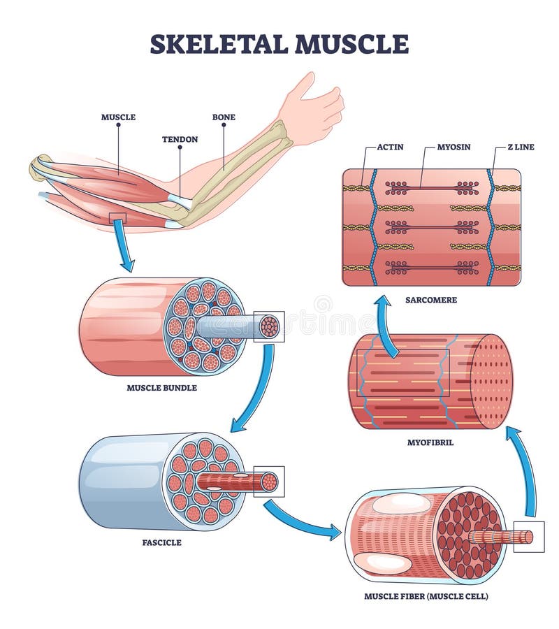
Myofibril - Wikipedia A myofibril (also known as a muscle fibril or sarcostyle) is a basic rod-like organelle of a muscle cell. Skeletal muscles are composed of long, tubular cells known as muscle fibers, and these cells contain many chains of myofibrils. Each myofibril has a diameter of 1–2 micrometres. They are created during embryonic development in a process known as myogenesis.

Histology of muscle | Body muscle anatomy, Skeletal muscle ...
sarcomere labeled diagram - Quizlet only myosin. actin. thin filaments. z line. end of sarcomere. cross bridges. when the myosin heads interact with thin filaments during a contraction. Upgrade to remove ads. Only $2.99/month.

Label the following in a diagram of a skeletal muscle fiber ...
Diagram Of Sarcomere The sarcomere is the contractile unit of muscle. This means it is the part of muscle Diagram of the Sarcomere. Source: . A dark stripe called a Z disc marks the ends of one sarcomere and the beginning Look at the diagram above and realize what happens as a muscle contracts.Mass Haul Diagram Explained. Whirlpool Duet Dryer Parts Diagram.
Sarcomere - Definition, Structure, Function and Quiz ...
Sarcomere model Diagram | Quizlet sarcomere contraction Actin filament Thin filament made mostly of actin subunits plus proteins troponin and tropomyosin Myosin head Tropomyosin binding site for myosin head Troponin regulatory protein with 3 polypeptide subunits. One subunit binds to actin, one to tropomyosin and another to calcium Triad

Sarcomere - Wikipedia
Sarcomere - Muscle Contraction - SmartDraw Muscle Contraction Skeletal muscle, also called striated muscle tissue, is made up of a series of sarcomeres A sarcomere consists of myosin and actin filaments which overlap upon contraction Myosin bonds with actin to ratchet the tropomyosin down the length of the myosin. Sarcomere Actin filament Troponin Tropomyosin Myosin filament
247 Sarcomere Images, Stock Photos & Vectors | Shutterstock
Sarcomere - Definition, Structure, Function and Quiz ... Mar 28, 2019 · A sarcomere is the functional unit of striated muscle. This means it is the most basic unit that makes up our skeletal muscle. Skeletal muscle is the muscle type that initiates all of our voluntary movement. Herein lies the sarcomere’s main purpose. Sarcomeres are able to initiate large, sweeping movement by contracting in unison.

Schematic representation of a sarcomere. The thick and thin ...
Two-Photon Excitation Microscopy for the Study of Living Cells … Jabłoński (energy-level) diagram of conventional one-photon excitation (left) and nonlinear two-photon excitation (right) of fluorescence. In each case, the absorption of photon(s) generates an excited state from which the molecule can relax by emitting a fluorescent photon. Thus, the path to the excited state follows a different path under either one- or two-photon absorption, leading …

Nebulin specifies thin-filament length and the level of ...
Sarcomere Labeling Quiz - PurposeGames.com About this Quiz This is an online quiz called Sarcomere Labeling There is a printable worksheet available for download here so you can take the quiz with pen and paper. Your Skills & Rank Total Points 0 Get started! Today's Rank -- 0 Today 's Points One of us! Game Points 8 You need to get 100% to score the 8 points available

Describe the structure of a sarcomere with a labelled diagram ...
Phytohormones (Plant hormones) - The Biology Notes 03.01.2021 · Types of Plant Cell- Definition, Structure, Functions, Labeled Diagram; Progress in Industrialization. For the industrial production of plant hormones such as GAs and ABA, several processes using fungal fermentation were successfully set up in China. As early as the 1950s–1960s, the solid-state fermentation of GAs has been applied. Compared with GAs, the …

Identifying the Z Line in the Sarcomere
Solved 12. Label the following diagram of a sarcomere | Chegg.com Label the following diagram of a sarcomere (filaments, bands, lines, etc., ...) ***Which regions shorten during contraction? 13. Describe four actions of ATP in the excitation-contraction coupling in a skeletal muscle fiber. 12-1. 12-2 12-3. 12-4. 14. The neuron and all the muscle fibers it excites are referred to as a 15.
Muscle fibre/sarcomere labelling - Labelled diagram
Question Video: Identifying the Z Line in the Sarcomere | Nagwa Two successive Z lines mark the boundary of each sarcomere. The A bands, labeled here with the letter Z, are the regions of the sarcomere that do contain the thicker, darker myosin filaments. And so they are sometimes called the dark bands. The outer edges of the A bands are darkest as they contain both actin and myosin filaments overlapping.

Striated muscle sarcomere. a Schematic diagram showing the ...
Structure and Function of the Skeletal Muscle Extracellular Matrix 01.09.2011 · Furthermore, multiple reaction monitoring may be used in the future to quantify proteins from labeled cell types in muscle. 107 This method can detect and quantify peptides in multiplex format using a triple quadrupole instrument. An advantage to this method is that it might be used to identify protein signals in fibrotic muscle that cause cells to overexpress collagen.
Sarcomere Labeled | EdrawMax Template
Home Page: Journal of Hand Surgery 26.05.2022 · The Journal of Hand Surgery publishes original, peer-reviewed articles related to the pathophysiology, diagnosis, and treatment of diseases and conditions of the upper extremity; these include both clinical and basic science studies, along with case reports.Special features include Review Articles (including Current Concepts and The Hand Surgery Landscape), …

Sarcomeres | BioNinja

RAPID - The Sarcomere The smallest functioning unit of a ...
Sarcomere Muscular Biology Scheme Vector Illustration Stock ...

Sarcomere location of MyBP-C. The electron micrograph was ...

Sarcomeric organization and MyBP-C. (A) Cardiac muscle ...

File:Sarcomere.svg - Wikimedia Commons
Draw the diagram of a sarcomere of skeletal muscle showing ...

5 The sliding filament model of muscle contraction. When a ...

191 Sarcomere Stock Photos, Pictures & Royalty-Free Images ...

Structure and position of titin in a sarcomere. | Human ...
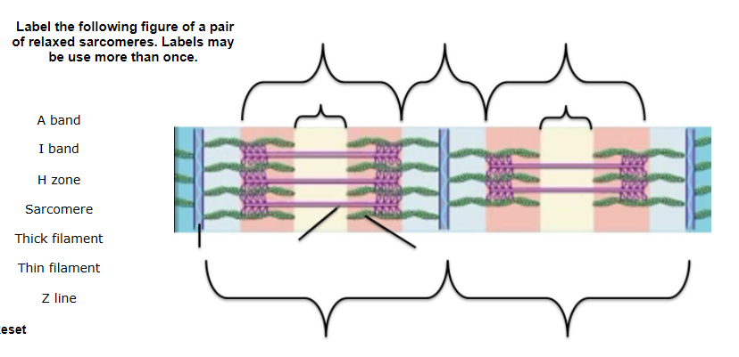
Solved Label the following figure of a pair of relaxed ...
Skeletal Muscle | Anatomy and Physiology I | | Course Hero
Sarcomere Contraction - Process Of Muscle Contraction With ...

Sarcomere - an overview | ScienceDirect Topics

Sarcomere Labeling Diagram | Quizlet

Layout of titin and other proteins in skeletal muscle ...

Schematic diagram of a muscle sarcomere. The isotropic and ...

10.2 Skeletal Muscle – Anatomy & Physiology
Sarcomere Muscular Biology Scheme Vector Illustration Stock ...
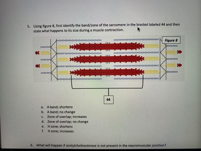
Solved 5) Using figure 8, first identify the band/zone of ...

Illustrations of (a) the sarcomere structure and (b) the ...

Sarcomere Diagram | Quizlet

UNIT 5: Label the parts of the Sarcomere Flashcards | Quizlet
Sarcomere Structure : Mnemonic | Epomedicine
Post a Comment for "40 sarcomere diagram labeled"