44 thigh muscle chart
Posterior thigh muscles - Hamstrings | Kenhub The hamstring muscles, or simply the hamstrings, are a group of three long muscles located in the posterior compartment of the thigh, shaping up the surface anatomy of this region. These muscles are the biceps femoris, semimembranosus and semitendinosus muscles. The hamstrings are closely related to each other as they share a common origin ... Leg Muscle Anatomy: Function & Facts | Openfit This muscle is just below the pectineus. Adductor magnus. One of the largest muscles in the body, this attaches along the linea aspera, a line running vertically along the inside of your thighbone. Adductor brevis and adductor longus. These adductors trace a similar path, reinforcing the action of the broad central portion of the adductor magnus.
Muscle Charts of the Human Body — PT Direct For your reference value these charts show the major superficial and deep muscles of the human body. Superficial and deep anterior muscles of upper body Superficial and deep posterior muscles of upper body
Thigh muscle chart
Univ of Michigan - Gross Anatomy - Muscles Tables the erector spinae m. is separated into 3 columns of muscle: iliocostalis laterally, longissimus in an intermediate position and spinalis medially; each of these columns has multiple named parts. iliocostalis. iliac crest and sacrum. angles of the ribs. extends and laterally bends the trunk and neck. Leg Muscle Diagram Pictures, Images and Stock Photos Calf Muscles - Anatomy Muscles isolated on white - 3D illustration Human thigh muscle anatomy for the education. Muscular system legs Human muscular system of legs in back view. Gluteus medius, gluteus maximus gastrocnemius and other muscles. Pelvis, leg and hip bones skeleton poster. Bodybuilding and strong body vector illustration Muscle Measurement Chart for Men - Dennis B. Weis the following 10 exercises and know for sure. BARBELL BACK SQUAT SUPINE BARBELL BENCH PRESS CONVENTIONAL DEADLIFT ONE-DUMBBELL PRESS RATE YOUR LIFT: DUMBBELL PRESS BARBELL WALL CURL BARBELL REVERSE CURLS WEIGHTED CHINS WEIGHTED DIPS ONE DUMBBELL TRICEPS EXTENSION WEIGHTED PUSHUPS ANOTHER STRENGTH STANDARD CHART FOR MEN STRENGTH STANDARDS FOR WOMEN
Thigh muscle chart. Thigh Anatomy, Diagram & Pictures | Body Maps - Healthline Muscles in the medial thigh help to bring the thigh toward the midline of the body and rotate it. These muscles are the adductor longus , adductor brevis , adductor magnus , gracilis, and the... Leg Muscles: Anatomy and Function - Cleveland Clinic The muscles in your upper and lower legs work together to help you move, support your body's weight and allow you to have good posture. They enable you to do big movements, like running and jumping. They also help you with small movements, like wiggling your toes. Leg muscle strains are common, especially in the hamstrings, quads and groin. Thigh Muscles Diagram, Pictures, List of Actions - Healthhype.com Muscle Pectinues Actions Adduction and flexion of the thigh Assists with medial rotation of the thigh Muscles Iliopsoas (psoas major, psoas minor, iliacus) Actions Flexion of the thigh at the hip joint Stabilizing the hip joint Muscle Sartorius Actions Abducts and flexes thigh at the hip joint Laterally rotates the the thigh at the hip joint Muscles of the Medial Thigh - TeachMeAnatomy The muscles in the medial compartment of the thigh are collectively known as the hip adductors. There are five muscles in this group; gracilis, obturator externus, adductor brevis, adductor longus and adductor magnus. All the medial thigh muscles are innervated by the obturator nerve, which arises from the lumbar plexus.
Hip and thigh muscles: Anatomy and functions | Kenhub The gluteal muscles can be divided into two main groups: Large and superficial muscles which mainly abduct and extend the thigh at the hip joint. These are the gluteus maximus, gluteus medius, gluteus minimus, and tensor fasciae latae. Small and deep muscles which mainly externally rotate the thigh at the hip joint and stabilize the pelvis. Thigh muscles Diagram | Quizlet A: flex leg and extend thigh gluteus maximus origin insertion action O: iliac crest, sacrum, coccyx I: linea aspera of femur, iliotibial band A: extend and laterally rotate thigh gluteus medius and minimus origin insertion action O: lateral, posterior surface of iliac I: greater trochanter of femur A: abduct and medially rotate thigh Muscles of the Thigh - Anterior - Medial - TeachMeAnatomy Muscles of the Thigh; Muscles in the Anterior Compartment of the Thigh. View Article. Muscles in the Medial Compartment of the Thigh. View Article. Muscles in the Posterior Compartment of the Thigh. View Article. Anatomy Video Lectures. START NOW FOR FREE. TeachMe Anatomy. Part of the TeachMe Series. Leg Muscles: Thigh and Calf Muscles, and Causes of Pain There are two main muscle groups in your upper leg. They include: Your quadriceps. This muscle group consists of four muscles in the front of your thigh which are among the strongest and largest...
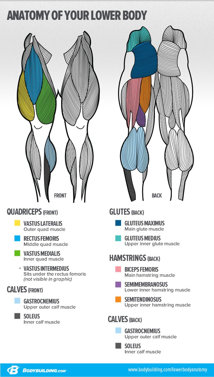
Thigh Muscles: Anatomy, Function & Common Conditions Quadriceps include four large muscles located in the front of the thigh: vastus lateralis, vastus medialis, vastus intermedius, and rectus femoris. They start at the pelvis (hip bone) and femur (thigh bone) and extend down to the patella (kneecap) and tibia (shin bone). Sartorius muscle is a long, thin muscle — the longest in the human body. Thigh Muscle Diagram | Leg muscles diagram, Muscle diagram, Leg muscles ... If so, these resistance band leg stretches are going to benefit you greatly. These gentle lower body stretches will help you relieve sore and tight leg muscles - calves, hamstrings, quads, glutes & hips - allowing you to speed up the recovery process and get back to moving normally. Upper limb arm forearm muscles actions, Blood supply ... Muscles of the Hip - Anatomy Pictures and Information The adductor muscle group, also known as the groin muscles, is a group located on the medial side of the thigh. These muscles move the thigh toward the body's midline. Included in this group are the adductor longus, adductor brevis, adductor magnus, pectineus, and gracilis muscles. Leg Muscle Diagram : Skeletal Muscle Review (With images) | Lower leg ... Leg muscles diagram labeled : The sacrum bone is almost always noticeable, no matter what the body type the accompanying muscle diagram reveals the position of the muscles of the lower legs in this pose. Human leg muscles diagram human leg muscle diagram anatomy body diagram. More commonly referred to as the 'quads,' no workout is complete ...
7 Lessons That Will Transform Your Legs! | Bodybuilding.com
Leg Muscles Anatomy, Function & Diagram | Body Maps Gastrocnemius (calf muscle): One of the large muscles of the leg, it connects to the heel. It flexes and extends the foot, ankle, and knee. Soleus: This muscle extends from the back of the knee to ...
Anatomy of the Quadriceps Muscles - Vastus Medialis, Vastus Intermedius ...
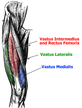
Muscles of the hips and thighs | Human Anatomy and Physiology Lab (BSB ... Figure 9-8. The superficial muscles of the thigh. Figure 9-9. The quadriceps group of four muscles. The view on the left has the rectus femoris cut away to show the vastus intermedius which is below it. The sartorius muscle is a distinctively long and thin muscle that crosses the thigh diagonally. It is visible in Figure 9-8.

Anatomy of the Knee | Health Life Media
Chart of Major Muscles on the Front of the Body with Labels A muscle of the medial thigh that originates on the pubis. It inserts onto the linea aspera of the femur. It adducts, flexes, and rotates the thigh medially. It is controlled by the obturator nerve. It pulls the leg toward the body's midline (i.e. adduction) Biceps brachii An upper arm muscle composed of 2 parts, a long head and a short head.

Muscles of the lower leg and foot | Human Anatomy and Physiology Lab ...
Average Thigh Circumference and Size in Males and Females Additionally, you'll discover how to measure your thighs the right way so that you can get the most accurate measurement and see how your legs stack up. 14 inch thighs 17 inch thighs 18 inch thighs 19 inch thighs 20 inch thighs 21 inch thighs 22 inch thighs 23 inch thighs 24 inch thighs 25 inch thighs 26 inch thighs 27 inch thighs 28 inch thighs

Tensor Fasciae Latae
Anatomy Tables - Muscles of the Lower Limb anterior tibial a. one of the muscles involved in anterior compartment syndrome. fibularis (peroneus) brevis. lower one third of the lateral surface of the fibula. tuberosity of the base of the 5th metatarsal. extends (plantar flexes) and everts the foot. superficial fibular (peroneal) nerve. fibular (peroneal) a.

Leg Muscles Diagram and the Cure!
Muscle Charts - Anatomy Guy Muscle Charts; Fun Videos; E-Learning. Curriculum; My Courses; Navigation best viewed on larger screens. Try using search on phones and tablets. ... Anterior Thigh Medial Thigh Posterior Thigh Anterior Leg Lateral Leg Posterior Leg Foot. Visceral. Curriculum Register / Sign In. Tools. Quiz Builder Muscle Charts. Supported By.

Top 5 Glute Stretches to Unlock a Bigger & Rounder Butt | Viva La Vibes
Muscle anatomy reference charts: Free PDF download | Kenhub Lower limb (free PDF download) This muscle chart eBook covers the following regions: Inner hip & gluteal muscles. Anterior, medical and posterior thigh muscles. Anterior, lateral and posterior leg muscles. Dorsal and plantar foot muscles. This eBook contains high-quality illustrations and validated information about each muscle.

myotomes chart - Google Search | Physical therapy assistant, Physical ...
Leg Muscle Chart_Spr20.pdf - Origin Iliopsoas Ad duct ors... Unformatted text preview: Origin Iliopsoas Ad duct ors Medial hip muscles: Adduction of Hip/leg Adductors pubis Gracilis Anterior hipmuscles: prime movers of flexion Iliacus Iliac crest Psoas major lumbar vertebrae Posterior hip muscles: Abduction Piriformis sacrum (deep!) Gluteus medius lateral ilium Gluteus maximus ilium, sacrum, coccyx Tensor fasciae latae iliac crest, ASIS Iliotibial tract ...

Pin on carn, jeans y pits⚛
Muscles that act on the Anterior Thigh • Anatomy & Function The quadriceps muscle is the largest muscle of the human body, and one of the prime movers of the leg. Learn more about the insertion, origin, innervation, and action of this muscle in this interactive and illustrated tutorial. Learn anatomy faster and. remember everything you learn. Start Now.
Post a Comment for "44 thigh muscle chart"