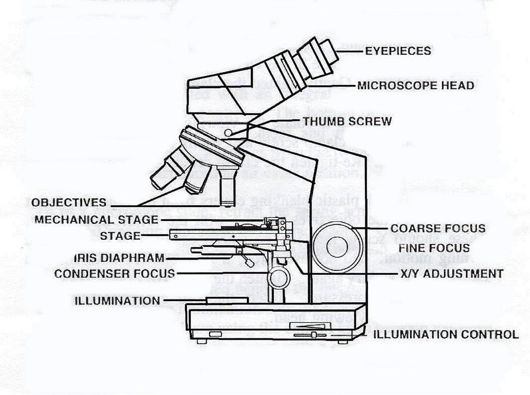
41 label the image of a compound light microscope
Compound Microscope: Definition, Diagram, Parts, Uses, Working ... - BYJUS Compound microscope is a type of optical microscope that is used for obtaining a high-resolution image. There are more than two lenses in a compound microscope. Learn about the working principle, parts and uses of a compound microscope along with a labeled diagram here. Compound Microscope Parts, Functions, and Labeled Diagram Nov 18, 2020 · Compound Microscope Definitions for Labels. Eyepiece (ocular lens) with or without Pointer: The part that is looked through at the top of the compound microscope. Eyepieces typically have a magnification between 5x & 30x. Monocular or Binocular Head: Structural support that holds & connects the eyepieces to the objective lenses.
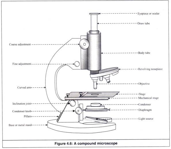
Parts of a microscope with functions and labeled diagram Q. List down the 18 parts of a Microscope. 1. Ocular Lens (Eye Piece) 2. Diopter Adjustment 3. Head 4. Nose Piece 5. Objective Lens 6. Arm (Carrying Handle) 7. Mechanical Stage 8. Stage Clip 9. Aperture 10. Diaphragm 11. Condenser 12. Coarse Adjustment 13. Fine Adjustment 14. Illuminator (Light Source) 15. Stage Controls 16. Base 17.

Label the image of a compound light microscope
Labelled Diagram of Compound Microscope - Biology Discussion The below mentioned article provides a labelled diagram of compound microscope. Part # 1. The Stand: The stand is made up of a heavy foot which carries a curved inclinable limb or arm bearing the body tube. The foot is generally horse shoe-shaped structure (Fig. 2) which rests on table top or any other surface on which the microscope in kept. The diagram below represents a compound light microscope Several parts ... 74 The diagram below represents a compound light microscope. Several parts have been labeled. In order to make an image brighter, which labeled part of the microscope would most likely be adjusted? (1) A (3) ... Solved Microscope parts/labeling 9 Label the image of a - Chegg question: microscope parts/labeling 9 label the image of a compound light microscope using the terms provided. 1 points eyepiece eyepiece light source references references base arm slide holder arm stage mechanical stage fine adjustment knob power switch objectives mindeneer microscopy lab homework saved help save & exit submit eyepiece light …
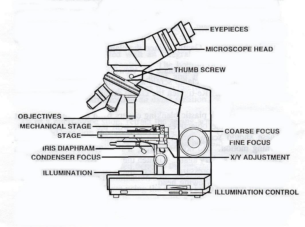
Label the image of a compound light microscope. Microscope With Labels clip art - Pinterest Download Clker's Microscope With Labels clip art and related images now. ... A diagram showing all of the parts of a compound light microscope. Microscope Parts and Functions With Labeled Diagram and ... Microscope Parts and Functions With Labeled Diagram and Functions How does a Compound Microscope Work?. Before exploring microscope parts and functions, you should probably understand that the compound light microscope is more complicated than just a microscope with more than one lens.. First, the purpose of a microscope is to magnify a small object or to magnify the fine details of a larger ... PDF The Compound Light Microscope The Compound Light Microscope TASK Refer to page 605 in your text to: 1. Name each of the structures described in the table to the right. 2. Match each structure to the letter in the diagram below. ** ALWAYS USE TWO HANDS TO CARRY A MICROSCOPE** Letter Structure Function joins body tube to base supports the entire microscope PDF Parts of the Light Microscope - Science Spot to SHARPEN the image G. BASE Supports the MICROSCOPE D. STAGE CLIPS HOLD the slide in place C. OBJECTIVE LENSES Magnification ranges from 10 X to 40 X F. LIGHT SOURCE Projects light UPWARDS through the diaphragm, the SPECIMEN, and the LENSES H. DIAPHRAGM Regulates the amount of LIGHT on the specimen E. STAGE Supports the SLIDE being viewed K. ARM
Parts of a Compound Microscope and Their Functions This number represents the compound microscope magnification power of the device. With the eyepiece, you may see a magnified image of the thing. Mirror: It is either fastened to the pillar or the lower end of the arm. On one side, it has a flat mirror, while on the other, it has a concave mirror. compound microscope parts (labeling) Flashcards | Quizlet Start studying compound microscope parts (labeling). Learn vocabulary, terms, and more with flashcards, games, and other study tools. ... light source of the microscope. what is 8? eyepiece (ocular lens) - magnifying piece that is looked into in order to see the specimen ... knob that brings the image to a sharper focus. what is 13? base - the ... Label the image to review the components of a compou... - Biology - Kunduz Question: Label the image to review the components of a compound light microscope. Nosepiece Arm k Mechanical stage ces Base Stage adjustm. Label the image to review the components of a compound light microscope. Nosepiece Arm k Mechanical stage ces Base Stage adjustment Ocular Fine focus Diaphragm Light source Coarse focus Objective lens Wames ... Bright-field microscope (Compound light microscope) - Diagram (Parts ... Bright-field Microscope. A bright-field microscope, also known as a compound light microscope is among the simplest of optical microscopes. Optical microscopes employ visible light and a series of lenses to magnify the specimen and view it in detail. A bright-field microscope uses light rays to create a dark image against a bright background ...
Compound Light Microscope: Everything You Need to Know A compound light microscope is a type of light microscope that uses a compound lens system, meaning, it operates through two sets of lenses to magnify the image of a specimen. It’s an upright microscope that produces a two-dimensional image and has a higher magnification than a stereoscopic microscope. Label the image of a compound light microscope Mar 9, 2022 — Label the image of a compound light microscope using the terms provided. Answer 1. eyepiece body body tube arm rotating nosepiece objectives ... Solved Label the image of a compound light microscope using ... Step-by-step answer. Who are the experts? Experts are tested by Chegg as specialists in their subject area. We review their content and use your feedback to keep the quality high. Transcribed image text: Label the image of a compound light microscope using the terms provided. Compound Microscope Labeled Diagram - Quizlet QUESTION. The total magnification of a specimen being viewed with a 10X ocular lens and a 40X objective lens is. 15 answers. QUESTION. a mosquito beats its wings up and down 600 times per second, which you hear as a very annoying 600 Hz sound. if the air outside is 20 C, how far would a sound wave travel between wing beats. 2 answers.
Free Microscope Drawing, Download Free Microscope Drawing png images ...
11 . O 2. Label the parts of the compound light micr... - Biology Label the parts of the compound light microscope on the diagram provided below (Figure 2-3). Names of these parts and their functions must be known to use the microscope correctly. ocular lens: remagnifies the image formed by the objective lens body tube: holds the lens system of the instrument arm: connects the body tube to the base; should be ...
Microscope light microscopy clipart simple pencil and inlor light 2 ...
Label Parts Compound Light Microscope Quiz | Shelly Lighting Compound Light Microscope Clipart 6 Best Photos Of Printable Microscope Worksheet Parts Microscope Bi 122 Biology Lab Series Microscopy Module A Study Of The Microscope And Its Functions With Labeled Diagram Compound Microscope Sketch At Paintingvalley Com Explore Learn About Microscopes With Fun Free Printables
Baker Demonstration Light Microscope with Mirror Reflecting Light ...
Compound Microscope- Definition, Labeled Diagram, Principle, Parts, Uses In order to ascertain the total magnification when viewing an image with a compound light microscope, take the power of the objective lens which is at 4x, 10x or 40x and multiply it by the power of the eyepiece which is typically 10x. Therefore, a 10x eyepiece used with a 40X objective lens will produce a magnification of 400X.

Diagram Of Microscope Example Binocular And Their Functions Compound ...
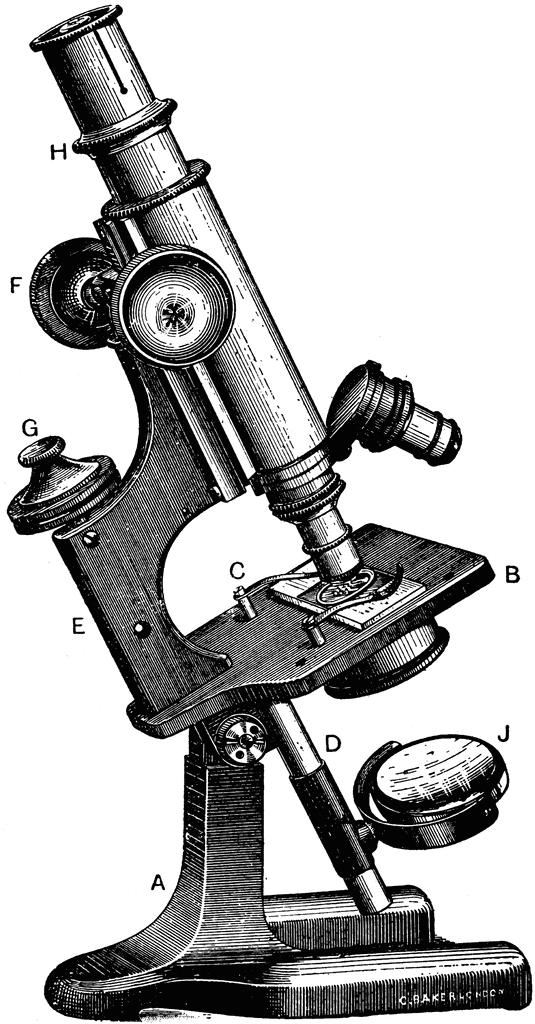
Label the above components of the compound light microscope A Ocular ... Label the above components of the compound light 4. Label the above components of the compound light microscope: A. Ocular lens (eyepiece) F. Rheostat B. Stage controls G. Spring loaded stage clip C. Coarse focus H. Illuminator D. Fine focus I. Iris diaphragm E. Objective lens 5.
Compound Microscope Drawing at PaintingValley.com | Explore collection ...
Compound Microscope - Types, Parts, Diagram, Functions and Uses Eyepiece/ocular lens - It is the part of the microscope that is looked through at the top. It comes with a magnification ranging between 5x and 30x. Image 3: The head connects the eyepiece to the objective lens. Picture Source: microscope.com. Head (monocular/binocular) - It is the structural support of the microscope.
PRACTICAL BOOKLET - BIOLOGY4ISC
Compound Microscope Parts – Labeled Diagram and their ... A compound microscope is the most common type of light (optical) microscopes. The term “compound” refers to the microscope having more than one lens. Basically, compound microscopes generate magnified images through an aligned pair of the objective lens and the ocular lens.

Compound light microscope | Microscopic, Microscope parts, Cool robots
Compound Microscope: Parts of Compound Microscope - BYJUS It is a U-shaped structure and supports the entire weight of the compound microscope. 2. Pillar. It is a vertical projection. This stands by resting on the base and supports the stage. 3. Arm. The entire microscope is handled by a strong and curved structure known as the arm. 4.

Image result for olympus CX31 microscope labeled | Microscope, Olympus ...
Label the image of a compound light microscope - Soetrust Which was the first cell viewed by the light microscope? Which of the following is true regarding the properties of… The compound below is treated with n-bromosuccinimide (nbs)…
Post a Comment for "41 label the image of a compound light microscope"